
Peroneal Nerve Entrapment Treatment: Clinic Care Before Surgery
Most cases of peroneal nerve entrapment respond to conservative care: activity changes, physical therapy, and bracing with an ankle-foot orthosis. Surgery is reserved for traumatic nerve transection, a compressing mass, rapidly worsening weakness, or persistent deficits after a monitored trial of nonoperative treatment. Recovery is often slow, and some people need an orthosis long after treatment ends.
TL;DR:
- An early normal EMG or nerve conduction study does not rule out entrapment; clinicians may repeat testing or compare both legs.
- Avoid leg crossing and pressure over the fibular head, use therapy and a neutral ankle brace, then reassess after several weeks to a few months.
- Surgery may be warranted for a cut nerve, compressing mass, rapidly worsening weakness, or deficits that persist despite a monitored conservative treatment trial.
- Nerves regenerate at roughly a millimeter a day, so recovery may take months or years; stretch, strengthen progressively, and use a brace to reduce falls.
Table of Contents
- What peroneal nerve entrapment is and what causes it
- Symptoms and when to see a clinician
- How peroneal entrapment is diagnosed
- Conservative treatment: activity change, therapy, bracing, and medication
- When surgery is considered and what the options look like
- Recovery and rehabilitation: what to expect and how to protect function
- How hands-on conservative care fits into a recovery plan
- Setting realistic expectations with patients
- How we help you manage peroneal nerve symptoms
- FAQ
- Sources
What peroneal nerve entrapment is and what causes it
The common peroneal (fibular) nerve wraps around the outside of the knee near the fibular head, then splits into superficial and deep branches that control ankle dorsiflexion, foot eversion, and sensation over the lower leg and top of the foot. When this nerve gets compressed or stretched, those functions falter.
Common causes include:
- Direct pressure at the fibular head from prolonged kneeling, squatting, or tight casts
- Habitual leg crossing, which compresses the nerve against the bone
- Trauma, including fractures near the knee or penetrating injuries
- Sports-related swelling or muscle entrapment in athletes with heavy lower-leg use
When the nerve can’t fire properly, the foot can’t lift or roll outward normally, which produces the classic foot drop and numbness pattern described below.
Symptoms and when to see a clinician
The hallmark sign is foot drop: the front of the foot catches or drags, causing tripping, especially on stairs or uneven ground. Weakness in dorsiflexion and eversion, along with numbness over the outer shin and top of the foot, often accompany it. Many people notice tripping before they notice any pain at all, and a painless presentation does not mean the problem is minor.
Seek prompt evaluation if you notice:
- Weakness that is getting worse over days rather than staying stable
- An open wound, cut, or penetrating injury near the outer knee
- Severe or spreading numbness alongside the weakness
- No improvement at all after several weeks of self-care
How peroneal entrapment is diagnosed
Diagnosis starts with a focused exam: manual strength testing of the muscles that lift and evert the foot, tapping over the fibular head to check for a Tinel sign, and watching gait for the telltale foot slap or high-stepping pattern that compensates for drop.
Electrodiagnostic testing, EMG combined with nerve conduction studies, helps pin down the location, severity, and likely prognosis of the nerve injury, but timing matters. EMG and nerve conduction studies can miss a lesion if done too early or without comparing both legs, so a normal early result doesn’t rule out entrapment.
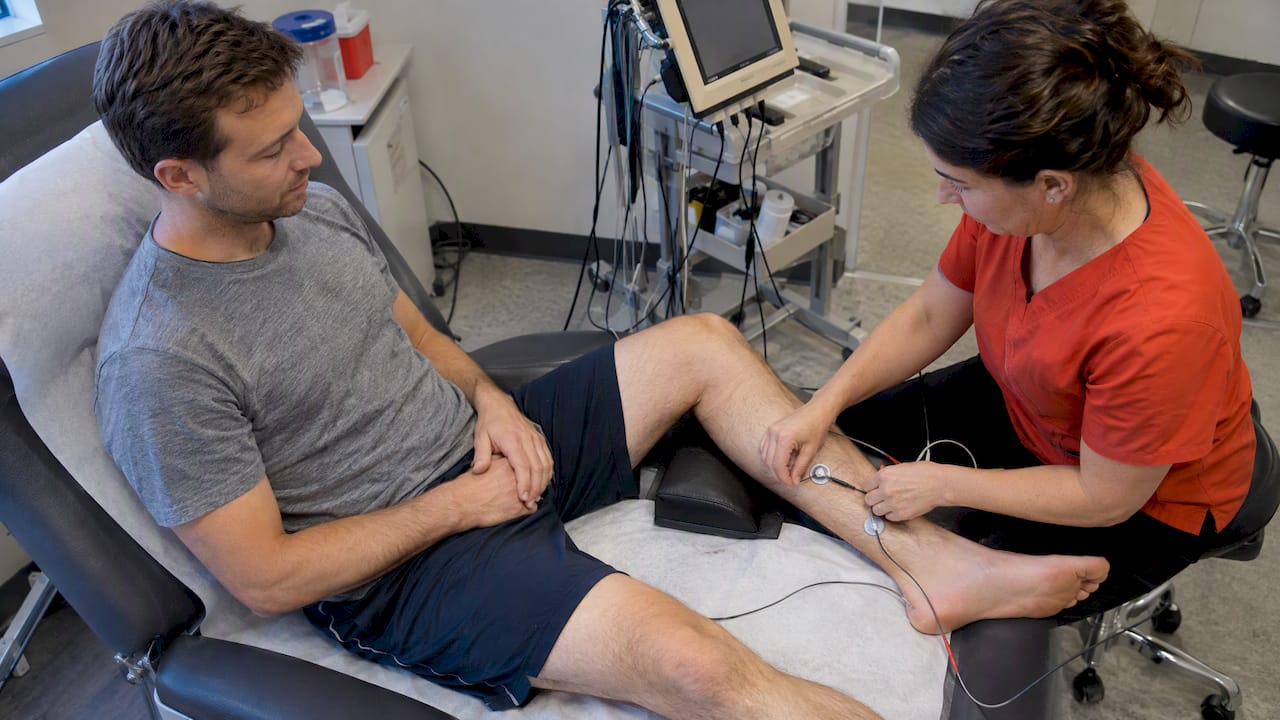
Some patients with suspected peroneal neuropathy may have normal early electrodiagnostic findings, which is why repeat testing or clinical correlation often matters more than a single test result.
Ultrasound or MRI gets added when a clinician suspects a mass, cyst, or structural compression, and these images become especially useful for planning surgery if that path is being considered.
Conservative treatment: activity change, therapy, bracing, and medication
For most people, first-line care means removing the source of compression and rebuilding strength around it. That starts with avoiding leg crossing, padding pressure points, and adjusting work or sports habits that load the fibular head repeatedly.
Physical therapy typically includes:
- Strengthening the tibialis anterior and evertor muscles to rebuild dorsiflexion
- Range-of-motion and proprioception drills to protect the ankle joint
- Gait retraining, sometimes paired with electrical stimulation to support weak muscles
Ankle-foot orthoses prevent tripping by holding the foot in a neutral position during the swing phase of walking, and newer carbon-fiber designs are lighter and return more energy at push-off than older plastic braces, which many patients find easier to tolerate for daily wear.
Pro Tip: Track your dorsiflexion strength and tripping frequency weekly; a clear trend, in either direction, gives your clinician the objective data needed to decide whether conservative care is working.
Medications for nerve-related pain, such as gabapentin or certain SNRIs and TCAs, are sometimes added when discomfort is significant. For superficial entrapments that resist standard care, image-guided procedures like ultrasound-guided pulsed radiofrequency have been reported to ease symptoms in carefully selected cases, including a documented case involving a professional athlete. Most clinicians give conservative measures a trial of several weeks to a few months before revisiting next steps. If you’re managing a related tendon issue alongside nerve symptoms, our guide on peroneal tendonitis treatment walks through a similar progressive approach.
When surgery is considered and what the options look like
Surgery enters the conversation under specific circumstances: a confirmed nerve transection, a mass pressing on the nerve, motor loss that’s worsening quickly, or no meaningful improvement after a fair trial of conservative care. Conservative management remains the initial standard for most cases without a traumatic laceration, which is why surgery is typically the second step, not the first.
Surgical options include:
- Decompression or neurolysis to free the nerve at the fibular head
- Direct repair or nerve grafting when the nerve has been cut or severely damaged
- Nerve transfers to reroute signal from a healthy donor nerve
- Tendon transfers as a salvage option for long-standing, chronic foot drop
Outcomes vary by technique, timing, and injury mechanism. A surgical series reported that a notable proportion of patients achieved antigravity dorsiflexion strength or better after surgery, with outcomes varying widely depending on technique and patient selection, according to the Acta Neurochirurgica surgical series. Earlier surgery after injury tends to track with better recovery, and some patients still need an orthosis after the operation.
Recovery and rehabilitation: what to expect and how to protect function
Peripheral nerves regenerate slowly, often at a rate of roughly a millimeter a day, which is why full recovery from peroneal entrapment can take months and sometimes stretches into years. Clinicians track progress through repeat strength testing and, when needed, follow-up electrodiagnostic studies.
Rehab priorities during that window:
- Daily passive stretching of the calf and ankle to prevent an equinovarus contracture from forming while muscles are weak
- Progressive strengthening as nerve signal returns, rather than waiting for full recovery to start
- Gait retraining with a physical therapist to build safer walking patterns
- Ongoing coordination between your orthotist and physical therapist so bracing adjusts as strength changes
Fall risk is real during recovery, so an assistive device or a well-fitted brace matters for daily safety, not just convenience. If progress stalls or reverses, that’s the signal to revisit imaging or a surgical opinion rather than wait indefinitely.
How hands-on conservative care fits into a recovery plan
Conservative management works best as a coordinated effort, and hands-on therapy has a role alongside bracing and traditional physical therapy. At our clinic, we apply the Fascial Distortion Model to address soft tissue restrictions around the fibular head and lower leg that can contribute to nerve irritation, paired with myofascial release and dry needling where appropriate.
This kind of manual work doesn’t replace electrodiagnostic testing or a surgical consult when either is warranted. Instead, it can be one piece of a broader team approach that includes physiatry, neurology, orthopedics, orthotists, and physical therapists, each covering the part of recovery suited to their training. When a patient’s presentation suggests a structural cause or a deficit that isn’t responding to hands-on work, we refer out for EMG or imaging rather than delaying that step.
Setting realistic expectations with patients
The patients who do best with peroneal nerve entrapment are the ones who treat gait safety and contracture prevention as daily habits, not occasional check-ins. Stretching and strengthening add up slowly, and skipping weeks sets recovery back further than most people expect.
I’d rather see a patient bring me a log of tripping frequency and strength changes than a vague sense that things feel “about the same.” That objective tracking is what lets us adjust a plan intelligently instead of guessing. If your symptoms are persisting past a few months or getting worse, an individualized assessment is worth prioritizing sooner rather than later.
— Chris
How we help you manage peroneal nerve symptoms
For those working through peroneal nerve symptoms, hands-on care can complement the strengthening, bracing, and medical follow-up your recovery needs. The approach often combines:
- Manual therapy techniques to address soft tissue tension around the knee and lower leg
- Dry needling for targeted muscle and nerve-adjacent irritation
- Focused shockwave therapy as an adjunctive option for stubborn symptoms
- Coordination with orthotists and referring physicians when bracing or further workup is needed
Your first visit includes a focused assessment of your movement pattern and soft tissue restrictions, and we’ll tell you plainly if your case needs electrodiagnostic testing or a surgical opinion before anything else. Book a visit with our team to get started.
This article is general information, not a substitute for advice from a qualified doctor. Consult a qualified healthcare professional about your own circumstances before acting on anything here.
FAQ
What exercises help strengthen the peroneal nerve pathway?
Exercises target the muscles the nerve controls rather than the nerve itself, focusing on ankle dorsiflexion and eversion strengthening, resistance band work, and balance drills. A structured, progressive program, like the ankle-strengthening guidance from NRTH, can help rebuild stability as nerve function returns.
What helps manage nerve pain day to day?
Protecting the fibular head from pressure, avoiding prolonged leg crossing, and pacing activity to avoid flare-ups are practical daily steps. Medications such as gabapentin or certain antidepressant classes are sometimes used for persistent nerve pain alongside physical therapy.
What is the path of the peroneal nerve in the leg?
The common peroneal nerve runs behind the knee, wraps around the outer side of the fibula near the fibular head, and then splits into superficial and deep branches that travel down the lower leg to control foot movement and sensation on the top of the foot.
Can crossing your legs cause peroneal nerve damage?
Yes, habitually crossing your legs can compress the peroneal nerve against the fibular head, and repeated or prolonged compression is a recognized cause of temporary or, in persistent cases, lasting nerve symptoms. Changing position regularly reduces this risk.
How long does recovery from peroneal nerve entrapment take?
Recovery often takes months and sometimes extends into years because nerve tissue regenerates slowly. Some patients regain full strength with conservative care alone, while others need ongoing bracing even after treatment.
Sources
- Peroneal Mononeuropathy Treatment & Management: Medical Care, Surgical Care — Medscape
- Surgical management of peroneal nerve injuries — Acta Neurochirurgica (Springer)
- Electrodiagnostic Evaluation of Peroneal Neuropathy — StatPearls / NCBI Bookshelf
- Ultrasound-Guided Pulsed Radiofrequency Treatment for Superficial Peroneal Nerve Entrapment — PMC