

Fascia Pain in Plano: Self Care 2–3 Weeks, Then Seek Hands On Help
Fascia is a continuous, collagen-rich connective tissue network that wraps every muscle, organ, nerve and bone in your body, and it plays a bigger role in pain and stiffness than most people realize. When fascia loses its normal glide through inactivity, injury, or overuse, it can restrict range of motion and generate pain that mimics a muscle or joint problem. The encouraging part: a mix of manual therapy, targeted movement, and graded rehab helps most people restore that glide.
TL;DR:
- Fascia can become densified or develop adhesions due to dehydration or trauma, causing pain, restricted movement, and referred discomfort.
- Common fascia-related conditions include myofascial pain syndrome, plantar fasciitis, frozen shoulder, and post-surgical adhesions, often worsened by repetitive strain.
- Manual therapy, instrument-assisted mobilization, and self-care tools provide short-term relief, but long-term results require active rehabilitation and movement diversity.
- Signs of fascia pain include trigger points, loss of tissue glide, and persistent stiffness post-injury, while urgent symptoms involve worsening pain or swelling.
- Consistent movement variation, gradual loading, and professional assessment are key to maintaining fascia health and preventing re-tightening.
Table of Contents
- What Is Fascia? The Main Layers and Where They Show Up
- How Does Fascia Support Movement and Sensation?
- Why Does Fascia Cause Pain, and What Conditions Result?
- What Treatments Target Fascia, and How Strong Is the Evidence?
- Daily Habits That Keep Fascia Healthy
- When Should You See a Clinician About Fascia Pain?
- The Overlooked Piece of Fascia Care
- Getting Hands-On Help for Fascia-Related Pain in Plano
- Sources
What Is Fascia? The Main Layers and Where They Show Up
Fascia is a continuous, three-dimensional sheath of collagen and elastin that runs beneath your skin, wraps every muscle group, and suspends your internal organs. Anatomists generally break it into three main categories, each built a little differently and serving a different job in the body according to NCBI’s clinical anatomy resources on fascia.
Superficial fascia sits just under the skin and contains fat and loose connective tissue. It cushions and insulates, and it’s the layer that lets your skin slide over the muscle underneath when you move.
Deep fascia wraps muscles, bones, nerves, and blood vessels in a denser, more elastin-rich sheet. This is the layer most manual therapists target, because it’s the one that transmits force between muscle groups and can tighten into painful bands.
Visceral fascia suspends your organs in position and lets them expand and contract as you breathe, digest, and move. Some anatomists also separate out parietal fascia, which lines body cavities like the abdominal wall.
At the microscopic level, fascia is populated with fibroblasts and fasciacytes, cells that produce collagen and secrete hyaluronan, a lubricating molecule that lets fascial layers slide past each other without friction. When hyaluronan gets sticky or dehydrated, that glide disappears.
A few named examples show how this plays out clinically:
- Thoracolumbar fascia, the thick sheet spanning your lower back, links your legs, spine, and shoulders. It’s a frequent culprit in chronic low back pain that doesn’t trace to a disc or joint.
- Fascia lata, wrapping the thigh, gets blamed for iliotibial band syndrome in runners and cyclists when it thickens and loses pliability.
- Plantar fascia, the band running along the sole of the foot, is the tissue behind plantar fasciitis, one of the most common causes of heel pain.
How Does Fascia Support Movement and Sensation?
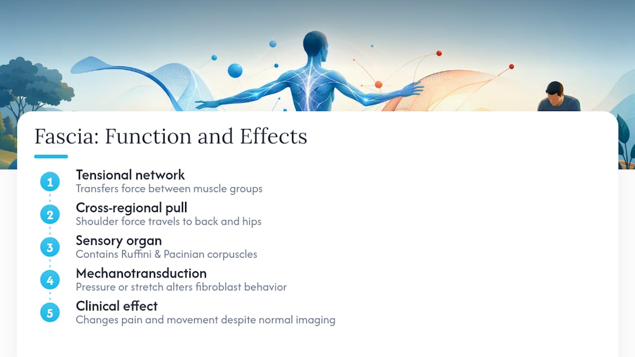
Fascia acts less like passive packing material and more like a body-wide tensional web that transmits mechanical force between muscle groups. When you reach overhead, the pull doesn’t stop at your shoulder. It travels through fascial connections down your back and into your hips, which is part of why a hip restriction can show up as shoulder pain.
Fascia also carries a dense supply of sensory receptors, including Ruffini and Pacinian corpuscles, giving it a genuine role in proprioception, your sense of where your body is in space. Research increasingly frames fascia as an organ contributing to both proprioception and pain signaling, not just a wrapper around the tissues that matter.
That sensory density explains a phenomenon patients often find confusing: fascial work can change how pain feels and how a joint moves, even when imaging shows no structural damage at all. The mechanism at play is called mechanotransduction. Applying steady pressure or stretch to fascia changes how fibroblasts behave and how the tissue matrix responds to load over time. It’s one reason clinicians treat fascia as an active, adaptable structure rather than a fixed piece of scaffolding.
Why Does Fascia Cause Pain, and What Conditions Result?
Fascia causes pain primarily through three mechanisms: densification, adhesion, and changes to hyaluronan. Densification happens when fascial layers thicken and lose their normal slide. Adhesions form when fascia sticks to adjacent tissue instead of gliding freely across it. And when hyaluronan dehydrates or becomes viscous, the layers that once moved smoothly start to catch and bind.
Several recognizable conditions trace back to these mechanisms:
- Myofascial pain syndrome produces trigger points, tight knots in fascia and muscle that refer pain to other areas of the body.
- Plantar fasciitis develops when repetitive strain thickens the plantar fascia, causing sharp heel pain, especially with the first steps in the morning.
- Adhesive capsulitis (frozen shoulder) involves fascial and capsular tightening around the shoulder joint that severely limits motion.
- Postsurgical adhesions form when fascial layers fail to glide properly after an operation. Over time these adhesions can vascularize and develop their own nerve supply, becoming an independent source of chronic pain long after the surgical site has healed.
- Compartment syndrome occurs when swelling inside a tight fascial compartment, often in the lower leg, cuts off blood flow and requires urgent care.
One detail worth remembering: manual fascial techniques typically apply sustained pressure for 15 to 120 seconds to encourage a release, a window that shows up consistently across clinical descriptions of the method.
Because fascia connects many structures, pain can refer to a location distant from its actual origin. A restriction in the thoracolumbar fascia may be perceived as a hip problem. A tight fascia lata can present similarly to knee pain. This referral pattern often leads to fascia-driven pain being misdiagnosed as a muscle strain or joint issue when the underlying cause is connective tissue.
What Treatments Target Fascia, and How Strong Is the Evidence?
Treatment options for fascia-related pain fall into three broad categories, and the right choice depends on the tissue, the timeline, and how the person responds to load.
- Manual myofascial release. A trained therapist applies slow, sustained pressure by hand, often for 15 to 120 seconds per point, until the tissue softens or releases. Studies suggest this approach can reduce chronic pain and improve function in select musculoskeletal conditions.
- Instrument-assisted soft tissue mobilization (IASTM). Clinicians use edged tools to apply targeted pressure across restricted fascia, often to break up adhesions that resist hand pressure alone.
- Device-based and self-care tools. Foam rollers, percussion devices, and shockwave therapy extend fascial treatment beyond the clinic. These are frequently used to maintain gains between professional sessions.
The evidence pattern across these methods is consistent: short-term improvements in pain and range of motion show up reliably, but long-term structural remodeling and modality-specific superiority remain inconsistent across studies. That’s not a reason to dismiss the approach. It’s a reason to treat fascial work as one piece of a bigger plan rather than a standalone fix.
The best-practice frame that’s emerged from that research treats fascial techniques as an adjunct to active rehabilitation, meaning exercise, motor control training, and progressive loading, rather than a substitute for it. Manual release opens a window of reduced pain and improved mobility; strengthening and movement retraining are what make that window last.
Aggressive fascial work isn’t right for every situation. Avoid deep pressure over acute inflammation, recent fractures, active infection, or suspected deep vein thrombosis, and always flag these to a provider before treatment begins.
Pro Tip: If a fascial release session leaves you sore for more than 48 hours or noticeably worse than before, that’s a signal the pressure or technique needs to be dialed back, not pushed through.
Daily Habits That Keep Fascia Healthy
Fascia stays supple through movement variety, not through any single stretch or gadget. Clinical guidance consistently points to frequent position changes, varied loading, and regular stretching as the foundation, since inactivity and repetitive strain are what dehydrate hyaluronan and cause stiffness in the first place.
A few practical habits go a long way:
- Change position every 30 to 45 minutes if you sit for long stretches at a desk.
- Rotate between different types of movement, walking, mobility drills, and strength work, instead of repeating the same motion pattern daily.
- If you use a foam roller or massage tool, move slowly, roughly one inch per second, to let the tissue’s viscoelastic response catch up rather than rolling fast and aggressive.
- Stop rolling or pressing if you feel sharp, shooting, or worsening pain rather than a dull, releasing ache.
Self-care has limits. If stiffness or pain persists for more than two to three weeks despite consistent effort, or if it’s interfering with sleep or daily function, that’s the point to bring in a professional assessment rather than keep troubleshooting alone.
When Should You See a Clinician About Fascia Pain?
Certain signs point toward fascial involvement specifically: palpable trigger points that reproduce pain when pressed, a noticeable loss of tissue glide in one area compared to the mirror side, and stiffness that persists well after a surgery, injury, or activity change resolves.
Clinicians assess this with hands-on palpation and movement testing, though imaging has real limits here. Ultrasound elastography can visualize some fascial stiffness, but it doesn’t capture the sensory and functional changes that drive most fascia-related pain.
Certain symptoms need urgent attention rather than a wait-and-see approach: rapidly worsening pain or swelling, numbness or weakness suggesting nerve involvement, tight and painful swelling that could signal compartment syndrome, or fever alongside localized pain that could point to infection.
The Overlooked Piece of Fascia Care
Most conventional advice on fascia treats it like a mechanical problem: find the tight spot, press on it, done. That framing undersells what the research actually shows. Fascia’s dense supply of sensory receptors means pain and stiffness are as much about nervous system input as they are about tissue texture, which is why two people with identical scan results can respond completely differently to the same release technique.
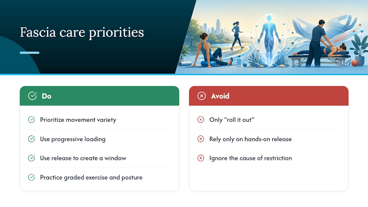
The bigger miss is sequencing. A lot of self-care advice stops at “roll it out” without addressing what caused the restriction to form. Fascia that tightens because of a strength deficit or a movement pattern flaw will just re-tighten after the next session unless something changes the load going through it.
If you take one thing from this article, prioritize movement variety and progressive loading over any single tool or technique. Foam rollers and hands-on release have a real, evidence-supported place, but they buy you a window of reduced pain and better mobility. What you do inside that window, the exercise, the posture correction, the graded return to activity, is what determines whether the improvement sticks.
— Chris
Getting Hands-On Help for Fascia-Related Pain in Plano
If foam rolling and stretching haven’t touched the stiffness, a hands-on evaluation often finds what self-care alone can’t reach. Thetxroom treats fascial restrictions directly, using the Fascial Distortion Model to locate specific patterns of fascial tightening and address them with targeted manual technique rather than generic stretching advice.

A first visit typically includes a movement assessment, hands-on evaluation of fascial glide in the affected area, and a treatment plan that combines manual work with active rehab and range-of-motion exercises. For pain that’s built up over time, Myofascial Acoustic Compression Therapy offers a device-assisted option alongside hands-on care. Many patients notice short-term relief early, with fuller, more durable change taking shape across a series of visits as rehab reinforces the manual work. If a persistent ache, a stubborn trigger point, or chronic back pain has been slowing you down, book an evaluation at Thetxroom and find out what’s actually driving it.
Sources
- PMC article on myofascial release techniques
- Frontiers in Physiology — Myofascial release and fascial-targeted interventions (2026 review)
- Cleveland Clinic — Fascia
This article is general information, not a substitute for advice from a qualified doctor. Consult a qualified healthcare professional about your own circumstances before acting on anything here.