
6–12 Week Plica Care for Clinicians: Clinic Tested Sequence to Avoid Surgery
Conservative management with activity modification and targeted physical therapy is the first-line treatment for plica syndrome, and it resolves symptoms in most patients. Corticosteroid injections serve as a short-term adjunct that can calm pain enough to let rehab progress. Arthroscopic plica resection is reserved for mechanical symptoms that persist despite an adequate conservative trial, typically several months.
TL;DR:
- Conservative treatment with activity modifications and physical therapy typically results in symptom resolution within six to twelve weeks, especially if compliance is maintained.
- Corticosteroid injections can provide short-term pain relief to enable rehabilitation but do not improve long-term outcomes without concurrent physiotherapy.
- Arthroscopic resection is reserved for cases with persistent mechanical symptoms after several months of conservative care and involves removing only the problematic plica tissue.
- Diagnosis relies heavily on clinical examination, with imaging mainly used to exclude other conditions and plan surgery when necessary.
- Long-term success largely depends on adherence to rehab protocols and the absence of cartilage damage, with isolated plica cases having better prognosis.
Table of Contents
- What Plica Syndrome Is and Why It Causes Pain
- Symptoms and Red Flags to Watch For
- Diagnosis: Clinical Exam, Imaging, and Diagnostic Injections
- Conservative Treatment: Activity Modification and a Structured Rehab Program
- Medications and Injections: Short-Term Relief and Limitations
- When Arthroscopic Resection Is Appropriate and What to Expect
- Recovery and Prognosis: Timelines and What Predicts Success
- Different Types of Plica and Why It Matters for Treatment
- Risks of Leaving Plica Syndrome Untreated
- Telling Plica Syndrome Apart From Other Knee Problems
- Long-Term Outcomes and Recurrence After Different Treatments
- A Clinician’s View on Getting This Right
- How We Support Conservative Knee Care
- FAQ
- Sources
What Plica Syndrome Is and Why It Causes Pain
A synovial plica is a fold of tissue left over from fetal knee development. Most people have at least one, and most never notice it. The medial plica, located along the inner side of the kneecap, is the one most often linked to pain because of its position and the friction it can create during bending and straightening.
Pain develops when a plica becomes irritated, thickened, or fibrotic. Repetitive flexion, direct trauma, or overuse can inflame the band, and once it thickens it can catch or rub against the femur with every step, a process doctors describe as impingement.
A few mechanisms explain most cases:
- Repetitive friction between the thickened plica and the femoral condyle during knee bending
- Altered patellofemoral tracking that increases pressure on the plica with each stride
- Fibrosis from a prior injury or chronic overuse that stiffens the normally pliable fold
Because the symptoms overlap heavily with meniscus tears and patellofemoral pain, accurate diagnosis matters as much as treatment. Treating the wrong structure delays recovery and can lead to unnecessary procedures.
Symptoms and Red Flags to Watch For
Plica syndrome usually announces itself with anterior or anteromedial knee pain that worsens with stairs, squatting, or sitting for long stretches with the knee bent. Many people notice a snapping or popping sensation along the inner kneecap, sometimes with a brief sensation of the knee catching before it releases, often called pseudolocking.
Typical features include:
- Dull, aching pain along the inside or front of the knee that builds with activity
- A snapping or popping sensation during flexion and extension
- Tenderness directly over the medial border of the patella
- Pain after prolonged sitting or repetitive stair climbing
Certain signs point away from plica syndrome and toward something that needs faster evaluation: true mechanical locking that does not release on its own, a large swollen joint, a feeling of the knee giving way, or fever and redness suggesting infection. Any of those warrants prompt medical assessment rather than a home exercise trial.
Pro Tip: Keep a simple log of what triggers your snapping or pain (stairs, squats, long drives) for a week before your appointment. That pattern often helps a clinician distinguish plica irritation from a meniscus or patellofemoral problem in a single visit.
Diagnosis: Clinical Exam, Imaging, and Diagnostic Injections
Diagnosing plica syndrome is mostly a clinical exercise. A thorough history and a set of provocative tests, feeling for a tender, cord-like band over the medial femoral condyle while bending and straightening the knee, often point to the diagnosis before any scan is ordered, according to a StatPearls overview of plica syndrome.
A practical diagnostic sequence looks like this:
- History and physical exam, focusing on pain location, snapping, and specific provocation maneuvers that reproduce symptoms.
- Imaging when needed, usually MRI or ultrasound, mainly to rule out a meniscus tear, cartilage damage, or other patellofemoral pathology rather than to confirm the plica itself, since clinical reviews note that imaging alone is not definitive.
- Diagnostic injection, where an intra-articular anesthetic can help separate pain coming from inside the joint from pain originating in surrounding soft tissue, though injecting the thin plica band directly is technically difficult and not routinely recommended without image guidance.
Imaging is also used to plan surgery when conservative care fails, since surgeons need to know whether any cartilage damage accompanies the plica before deciding how aggressive to be.
Conservative Treatment: Activity Modification and a Structured Rehab Program
Most people with plica syndrome improve with a disciplined conservative program built around three pillars: reducing the activities that provoke pain, restoring strength and control around the knee, and progressing gradually back to full function. Case series and cohort studies consistently show that structured rehabilitation leads to symptomatic improvement in most patients, with only a small fraction going on to surgery, according to cohort data on pathological synovial plicae.
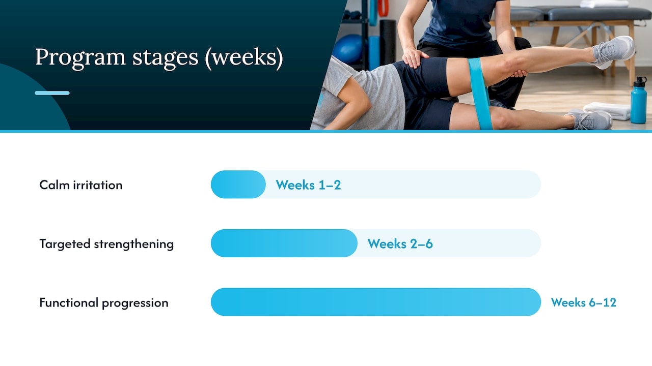
A typical program unfolds in stages:
- Weeks 1 to 2, focus on calming irritation: reduce deep squatting, stair repeats, and prolonged sitting with the knee bent; use ice after activity; begin gentle range-of-motion work without forcing painful end ranges.
- Weeks 2 to 6, add targeted strengthening: quadriceps activation with an emphasis on the vastus medialis obliquus (VMO), along with hip abductor and external rotator work to improve patellofemoral tracking.
- Weeks 6 to 12, progress to functional and neuromuscular control exercises, step-downs, single-leg balance work, and controlled lunges, while gradually reintroducing the activities that originally caused pain.
Stretching for the hamstrings and the IT band is typically woven throughout the program, since tightness in either structure increases lateral pull on the kneecap and indirectly aggravates the plica. Supervised sessions one or two times a week usually transition to a home program by the six-week mark, provided symptoms are trending down.
During an acute flare, open-chain loading exercises that place direct stress on a bent, painful knee, like deep leg extensions, are usually avoided until pain is controlled and tracking has improved. Our guide on patellar tendinopathy rehab progression covers similar loading principles for anterior knee pain that apply here.
The evidence base for this approach is solid. A randomized controlled trial comparing arthroscopic resection to structured physiotherapy found that surgery produced better Lysholm scores at two years, but both groups improved meaningfully in the short term, and patients who stuck with a disciplined program over time saw results that approached surgical outcomes. Compliance, more than any single exercise, is often what separates a good result from a frustrating one.
Pro Tip: Track your pain on a simple 0 to 10 scale before and after each session. If pain climbs above your baseline for more than a day, scale back the intensity rather than pushing through, since irritation that lingers tends to slow the whole program down.
Medications and Injections: Short-Term Relief and Limitations
Oral NSAIDs or acetaminophen are commonly used to manage pain during the early, most irritable phase of plica syndrome, paired with icing and simple activity pacing rather than complete rest.
When pain is intense enough to block progress in physical therapy, an intra-articular corticosteroid injection can help. These injections often reduce pain in the short term and can create a window where rehab becomes tolerable, but evidence for long-term benefit on their own is limited, so they work best as a bridge rather than a stand-alone fix.
A few practical points matter here:
- Injections are typically followed by a short period of relative rest, often a day or two, before resuming normal activity
- Repeated steroid injections are generally avoided because of concerns about cartilage and soft tissue effects over time
- Injections are usually skipped when there are signs of infection or when the diagnosis is still uncertain
- Regenerative options like platelet-rich plasma are sometimes discussed as alternatives; a comparison of corticosteroid versus PRP injections for joint pain outlines how the two differ in mechanism and expected timeline
The consistent theme across clinical guidance is that injections support rehab, they do not replace it.
When Arthroscopic Resection Is Appropriate and What to Expect
Surgery becomes a reasonable option when mechanical symptoms, true catching, persistent swelling, or pain that has not budged, continue despite an adequate conservative trial, commonly three to six months, or when a patient genuinely cannot comply with a structured rehab program.
What to know before considering it:
- Arthroscopic excision lets a surgeon directly visualize the plica, confirm the diagnosis, and remove only the pathological band rather than healthy synovial tissue, since clinical consensus warns against wide synovial excision to reduce the risk of postoperative fibrosis
- The same procedure allows a surgeon to address any cartilage lesions found at the same time, which is one reason imaging beforehand matters
- Outcomes are generally good when the plica is the only pathology present, but results are less predictable when cartilage damage coexists
- As with any arthroscopy, there are standard surgical risks, infection, stiffness, and a recovery period that requires its own rehabilitation to regain strength and motion
Surgery is a tool for a specific, well-defined problem, not a shortcut past a rehab program that has not yet been given a fair trial.
Recovery and Prognosis: Timelines and What Predicts Success
With conservative care, many patients notice meaningful improvement within a few weeks, and supervised programs typically run six to twelve weeks before transitioning fully to independent home exercise. Staying with a maintenance strengthening routine afterward appears to lower the odds of recurrence, though stopping cold once symptoms fade is a common reason pain returns.
After surgery, early range-of-motion work and gentle muscle activation begin almost immediately, followed by progressive strengthening over the following weeks. Our breakdown of recovery milestones for knee ligament injuries outlines a similar staged timeline that applies broadly to knee rehab, conservative or post-surgical.
A 2021 cohort review of plica resection outcomes found generally favorable short-term results, with more variable long-term outcomes when cartilage lesions were also present, according to outcome data on pathological synovial plica treatment. That single detail explains much of the variability patients encounter: an isolated plica with no other joint damage tends to do well, while a plica found alongside cartilage wear carries a less certain long-term outlook regardless of treatment path.
- Isolated plica without chondral damage: generally favorable outcomes with either conservative care or surgery
- Plica with coexisting cartilage damage: more variable long-term pain relief
- Strong rehab compliance: consistently associated with better and more durable results
Different Types of Plica and Why It Matters for Treatment
The knee can have up to four plicae, and which one is involved changes both symptoms and treatment emphasis. The suprapatellar plica sits above the kneecap and rarely causes trouble on its own. The infrapatellar plica runs below the kneecap toward the tibia and is usually incidental. The medial plica, the one most frequently implicated in plica syndrome, runs along the inner joint line and is positioned exactly where it can catch against the femoral condyle during flexion. A lateral plica is less common but can mimic lateral meniscus symptoms when irritated.
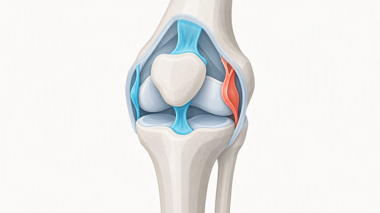
Knowing which plica is involved shapes the physical exam, since provocation tests are designed around the medial band specifically, and it shapes surgical planning, since a surgeon targeting the wrong fold during arthroscopy would miss the actual source of pain. It also explains why an incidental plica spotted on an MRI done for an unrelated reason should not automatically be blamed for a patient’s symptoms. Many people carry an infrapatellar or suprapatellar plica their entire lives without a single symptom, and clinical guidance specifically cautions against resecting an incidentally found plica that is not clearly the pain generator.
Risks of Leaving Plica Syndrome Untreated
Plica syndrome rarely threatens the joint the way a severe ligament or cartilage injury does, but ignoring persistent symptoms carries real downsides. Chronic friction between a thickened plica and the femoral condyle can gradually irritate the adjacent cartilage, and over months to years that irritation can contribute to early wear in that part of the joint.
Persistent pain also changes how people move. Favoring the knee to avoid the pinch of a snapping plica often leads to compensatory patterns, altered gait, reduced quadriceps activation, overloading the opposite leg, that create their own secondary problems, including hip or lower back strain.
There is also a functional cost that is easy to underestimate: ongoing anterior knee pain tends to shrink a person’s activity radius over time. Stairs get avoided, workouts get shortened, and the muscles that should be protecting the joint get weaker from disuse, which can make the original problem harder to reverse once someone finally seeks care. None of this means every case needs urgent intervention, but it does argue against letting symptoms linger indefinitely without at least starting a structured conservative program.
Telling Plica Syndrome Apart From Other Knee Problems
Plica syndrome shares real estate, both anatomically and symptomatically, with several other common knee conditions, which is exactly why a careful exam matters more than a quick guess.
A meniscus tear often causes joint-line tenderness and true locking that does not self-resolve, versus the pseudolocking and snapping more typical of plica irritation. Patellofemoral pain syndrome tends to produce a broader, less localized ache around or behind the kneecap rather than the discrete, cord-like tenderness found along the medial plica. Patellar tendinopathy concentrates pain at the bottom of the kneecap, worsened specifically by jumping or deep loading, rather than the snapping sensation characteristic of plica syndrome.
The exam itself relies on a combination of findings: a palpable, tender band over the medial femoral condyle, reproducible snapping with specific flexion and extension maneuvers, and the absence of joint-line tenderness that would point toward the meniscus instead. When the picture is unclear, imaging is brought in specifically to rule out these other diagnoses rather than to confirm the plica itself, since MRI findings of a plica are common even in people without symptoms. Getting this differentiation right the first time avoids months of treating the wrong structure.
Long-Term Outcomes and Recurrence After Different Treatments
For patients who complete a full conservative program, the long-term picture is generally encouraging, and recurrence is uncommon when a maintenance strengthening routine continues past the initial treatment window. The patients who tend to see symptoms return are usually the ones who stop all exercise once pain subsides rather than easing into a lower-intensity ongoing routine.
Surgical outcomes tell a more nuanced story. The two-year data from the randomized trial comparing resection to physiotherapy favored surgery on Lysholm scores, but both groups improved, and the gap narrowed for patients who stayed consistent with their exercise program over time, per the same RCT on arthroscopic resection versus structured physiotherapy. Longer-term cohort data following resection show generally good short-term relief, with outcomes becoming less predictable when cartilage damage was present alongside the plica.
The practical takeaway is that treatment choice matters less than most people assume, and consistency matters more. A conservative program carried through to completion, with ongoing maintenance work afterward, produces durable results for most people with an isolated plica. Surgery offers a faster, more reliable path for the subset whose mechanical symptoms do not respond to an adequate trial of rehab, but it does not erase the need for a solid post-operative strengthening program, and long-term pain relief after surgery still tracks closely with whether cartilage damage was present from the start.
A Clinician’s View on Getting This Right
Most frustration with plica syndrome comes from stopping rehab the moment pain eases rather than finishing the program. Adherence and correct technique matter more than which specific exercise you choose. We would escalate to imaging or a surgical referral only when mechanical symptoms persist past a genuine, well-executed conservative trial, not simply because progress feels slow in week two.
— Chris
How We Support Conservative Knee Care
Knee pain rarely responds to a single fix, which is why we build individualized, progressive plans rather than handing every patient the same sheet of exercises. For plica-related knee pain, we often combine hands-on soft tissue work using the Fascial Distortion Model with targeted rehab designed to restore normal tracking and loading tolerance around the joint.

Depending on what we find during your evaluation, your plan may include:
- Manual therapy techniques to address restricted soft tissue around the knee
- Dry needling for persistent muscular tension contributing to altered movement patterns
- Focused shockwave therapy as an adjunct for stubborn soft tissue irritation
- A progressive, individualized strengthening program adjusted session to session
A first visit starts with a hands-on evaluation of how your knee moves and loads, not a generic script. When findings suggest something beyond our scope, a torn meniscus or a case needing imaging, we refer out rather than treating blind. Book an evaluation at The Tx Room to get a plan built around your knee specifically.
This article is general information, not a substitute for advice from a qualified doctor. Consult a qualified healthcare professional about your own circumstances before acting on anything here.
FAQ
Does plica syndrome go away on its own?
Plica syndrome often improves with a structured conservative program rather than simple rest alone, and most patients see meaningful symptom relief within weeks of starting targeted activity modification and exercise. A small number of cases with persistent mechanical symptoms do not resolve without further treatment, including surgery in select situations.
How long does it take for an inflamed plica to heal?
Many people notice improvement within the first few weeks of a conservative program, with supervised rehab typically continuing for six to twelve weeks before transitioning to independent home exercise. Full recovery timelines vary depending on how long symptoms were present before treatment started and how closely the rehab program is followed.
What does an inflamed plica feel like?
An inflamed plica usually feels like a dull ache along the inner or front part of the knee, often paired with a snapping or catching sensation during bending and straightening. Tenderness is typically focused over a specific tender band on the inside of the kneecap rather than spread diffusely across the joint.
What are some common problems with the patella?
Beyond plica syndrome, common patella-related issues include patellofemoral pain syndrome, patellar tendinopathy, and tracking problems where the kneecap does not glide smoothly within its groove during movement. These conditions can produce overlapping symptoms, like anterior pain with stairs or squatting, which is why an accurate clinical exam matters before starting treatment.
Sources
- Synovial plica syndrome of the knee: a commonly overlooked cause of anterior knee pain – PMC
- Plica syndrome – StatPearls – NCBI Bookshelf