

Chondromalacia Patella Treatment: 6–12 Weeks of Evidence Based Rehab
Structured exercise therapy combined with patient education is the first-line, evidence-based treatment for chondromalacia patella, and it works better than doing nothing within the first three months. Skipping straight to injections or surgery is rarely the right first move. Escalation to injections or surgical evaluation makes sense only after a genuine, sustained rehab effort fails or imaging confirms a clear structural lesion, and most non-urgent cases deserve months of conservative care before elective surgery enters the conversation.
TL;DR:
- Most patients see short-term improvement within three months through targeted exercise therapy that strengthens hip and knee muscles.
- Conservative care, including education, activity modification, and orthoses, should be tried for several months before considering injections or surgery.
- Injections such as PRP or hyaluronic acid have limited evidence and are typically reserved for cases where rehab has failed.
- Surgery is usually only indicated after a documented failure of conservative treatment or if imaging reveals a clear structural lesion.
- Consistently tracking pain levels and adhering to a structured rehab plan are essential to avoid unnecessary escalation to invasive procedures.
Table of Contents
- Quick treatment summary: the main categories and their expected effects
- Exercise therapy: the rehab program that evidence supports
- Combined conservative measures: taping, braces and orthoses, and activity modification
- Medications and injection therapies: what the evidence says
- Surgery: indications, common procedures, and outcomes
- Diagnosis and staging: how tests influence treatment choices
- Recovery timeline, prognosis, and red flags
- How to choose the right treatment: a practical checklist and questions to ask
- How hands-on adjunctive care can fit into a rehab plan
- Role of physical therapy modalities (ultrasound, electrotherapy)
- Nutritional considerations relevant to cartilage health
- What the evidence really tells us about treating this knee condition
- The Tx Room: services that support chondromalacia patella rehab
- Sources
- FAQ
Quick treatment summary: the main categories and their expected effects
Chondromalacia patella treatment falls into a handful of buckets, and knowing where each one fits saves you from wasting time on the wrong approach first.
Conservative care is the foundation for nearly everyone. This includes patient education about the condition, a structured exercise program targeting the hip and knee, temporary activity modification, bracing or orthoses in select cases, and over-the-counter pain medication for flare-ups. According to StatPearls, nonsurgical therapy centered on targeted physiotherapy, activity modification, bracing, and orthotics is the recommended starting point, often continued for several months before surgery is considered.
Adjunctive measures sit on top of that foundation rather than replacing it. Patellar taping, foot orthoses, and hands-on soft tissue work can reduce pain enough to let you participate more fully in exercise. Injectable options, including platelet-rich plasma, hyaluronic acid, and mesenchymal stem cell injections, are considered when conservative care stalls, though the evidence behind them is mixed and far from settled.
- Education plus exercise: the combination most consistently linked to short-term improvement.
- Bracing, orthoses, and taping: symptom control tools that support participation in rehab rather than fix the underlying issue.
- Injections (PRP, HA, MSCs): selective options with inconsistent evidence, typically tried after rehab has been given a fair shot.
- Surgery: reserved for persistent, function-limiting cases or confirmed structural lesions that do not respond to nonoperative care.
A network meta-analysis of patellofemoral pain treatments found that education combined with a physical treatment was the approach most likely to help in the short term. Over longer-term follow-up, education alone may perform comparably if patients adhere to exercises. That single finding reshapes how you should think about this condition: the most important gains occur early and result from movement and education rather than procedures.
Surgery, by contrast, is selective. It is not the next logical step after a few weeks of home stretches; it is a decision made after a documented, adequate trial of rehab has failed or after imaging shows a lesion that explains persistent mechanical symptoms.
Exercise therapy: the rehab program that evidence supports
The rationale behind exercise therapy for chondromalacia patella is mechanical. The kneecap tracks in a groove formed by the thigh bone, and when the muscles that guide that tracking, particularly the vastus medialis obliquus and the hip external rotators and abductors, are weak or poorly coordinated, the patella rubs unevenly against the cartilage underneath it. Strengthening those muscles reduces that maltracking and the irritation that comes with it.
The evidence supports a specific shape for this training. A systematic review and meta-analysis found that knee-targeted exercise and combined hip-and-knee exercise programs both improved outcomes over shorter-term wait-and-see care, with combined programs generally edging out knee-only work. Clinical practice guidance from the APTA reinforces this, recommending exercise therapy that includes both hip and knee work rather than isolated quadriceps strengthening.
A practical program typically looks like this:
- Weeks 1 to 2: closed-chain short-arc quad sets, straight-leg raises, and gentle hip abduction and external rotation work, three to four sessions per week, focusing on pain-free range of motion.
- Weeks 3 to 6: progress to mini-squats, step-downs, and side-lying hip abduction with resistance bands, adding core stabilization work like planks and bird-dogs.
- Weeks 7 to 12: introduce single-leg squats, lateral step-ups, and controlled lunges, increasing load and range as pain allows, while monitoring for any increase in symptoms the following day.
- Beyond week 12: sport-specific or activity-specific drills for those returning to running, cycling, or jumping sports, layered on top of the maintained strength base.
Dosing generally follows a pattern of two to three sets of eight to fifteen repetitions, adjusted based on how the knee responds, and progression should be governed by symptoms rather than a fixed calendar. If an exercise causes pain that lingers for more than a day, it is a sign to regress rather than push through it.
Supervised physical therapy tends to produce more consistent adherence than a home program alone, largely because a therapist can adjust form and progression in real time. That said, a well-designed home program with clear milestones can work if you track your pain and function honestly and resist the urge to skip sessions once symptoms start improving, since early improvement is often the point where people quietly abandon the exercises that got them there.
Pro Tip: Keep a simple weekly log of your pain level (0 to 10) before and after each session. If pain trends downward over two to three weeks, keep progressing. If it plateaus or worsens, that is your cue to talk to a physical therapist before adding load.
Combined conservative measures: taping, braces and orthoses, and activity modification
Exercise alone is often not enough to get someone through the early, more painful weeks of rehab, which is where combined interventions earn their place. The same systematic review that supports exercise therapy also found that foot orthoses improved short-term outcomes compared to wait-and-see care, and the network meta-analysis on patellofemoral pain treatments identified education paired with a physical treatment, including taping or orthoses, as the most effective combination at three months.
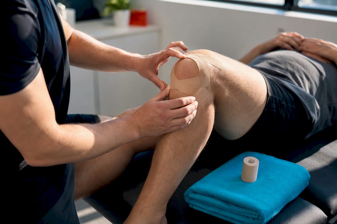
Foot orthoses may help people whose foot posture, such as excessive pronation, contributes to poor knee alignment during activity; clinical assessment can guide use. Patellar taping works differently: it is a short-term tool to reduce pain during activity, which in turn lets you tolerate more exercise volume during the early weeks of a program rather than avoiding movement out of discomfort.
Practical uses of these adjuncts include:
- Orthoses: worn during weight-bearing activity for those with pronounced foot pronation contributing to knee stress.
- Patellar taping: applied before workouts or activity to reduce pain enough to complete a rehab session.
- Bracing: occasionally used for added stability during return-to-sport phases, though it is not a substitute for strength work.
- Activity modification: temporarily reducing high-load activities like deep squats, stairs, or running downhill while symptoms are acute, then reintroducing them gradually as strength improves.
Return-to-activity guidance generally follows a stepwise approach: reduce or modify aggravating activities in the first two to four weeks, reintroduce them at lower intensity as pain allows, and only resume full training once you can perform functional tests, like single-leg squats or step-downs, without pain. Readers managing related knee issues may find the exercise and taping strategies covered for runner’s knee or patellar tendinopathy useful for cross-reference, since the rehab principles overlap significantly.
Medications and injection therapies: what the evidence says
Over-the-counter medications have a role, but it is a limited one. NSAIDs like ibuprofen or naproxen, and acetaminophen, can reduce pain during flare-ups and make it easier to participate in exercise, but neither changes the underlying cartilage or biomechanics driving the condition. They are best used short-term and alongside, not instead of, exercise therapy.
Injection therapies occupy a gray area. Platelet-rich plasma and mesenchymal stem cell injections have drawn attention as potential options when conservative care has not resolved symptoms. A review of emerging cell therapies notes that these approaches show promise for patellofemoral cartilage lesions, with some studies reporting symptomatic improvement, but the evidence remains preliminary and inconsistent, and structural cartilage repair is not reliably demonstrated. A partner resource on PRP in sports medicine echoes this caution, describing real benefits alongside meaningful limits that patients should understand before pursuing treatment.
Hyaluronic acid injections have limited and variable evidence for patellofemoral chondromalacia and are not standard recommendations.
- NSAIDs and acetaminophen: short-term symptom relief, no disease-modifying effect on cartilage.
- PRP and MSC injections: emerging options with inconsistent structural benefit; often not covered by insurance and can require multiple sessions.
- Hyaluronic acid: limited, variable evidence specific to patellofemoral chondromalacia.
- Cost consideration: biologic injections are frequently an out-of-pocket expense, and no source establishes a guaranteed long-term benefit that justifies the cost for every patient.
Emerging cell therapies, including autologous chondrocyte implantation and mesenchymal stem cell injections, show promise for patellofemoral cartilage lesions, but the evidence remains preliminary and inconsistent, according to a review of current and emerging treatments. That gap between early promise and proven benefit is exactly why these should be framed as experimental options, not a shortcut past rehab.
Surgery: indications, common procedures, and outcomes
Surgery becomes a reasonable conversation when persistent, function-limiting symptoms remain after a comprehensive, honestly-attempted rehab program, or when imaging and exam findings point to a specific mechanical or structural lesion that exercise cannot fix on its own. It is not typically the first, second, or even third option tried.
Common procedures include:
- Arthroscopic chondroplasty or debridement: smoothing damaged cartilage surfaces, usually the least invasive surgical option.
- Lateral release with medial reefing or realignment: addresses patellar tracking problems surgically when conservative measures have not corrected alignment.
- Tibial tubercle osteotomy: repositions the attachment point of the patellar tendon to change how forces load the kneecap.
- Patellofemoral arthroplasty: a partial knee replacement limited to the patellofemoral joint, reserved for more advanced, isolated cartilage loss.
- Partial or total patellectomy: rarely performed, generally a last resort for severe, refractory cases.
The evidence on outcomes tempers expectations considerably. A systematic review and meta-analysis on arthroscopic surgery for degenerative knee disease found that arthroscopic lavage and debridement provide, at best, very small short-term benefits over conservative care, with minimal or no meaningful advantage in the long run. That finding lines up with similar conclusions discussed in the context of hip labral tear conservative treatment, where surgical benefit over structured rehab is also modest for many cases. Patellofemoral arthroplasty has a narrower, more specific set of indications and mixed data on longer-term durability, making it a niche solution rather than a default one.
Recovery after these procedures varies widely by type, ranging from a few weeks of restricted weight-bearing after arthroscopy to several months of progressive rehab after realignment or arthroplasty procedures. Common complications to discuss with a surgeon include stiffness, persistent pain, and, in realignment procedures, the possibility of recurrent instability. These conversations belong with an orthopedic surgeon who can weigh your specific imaging and history, not a generic checklist.
Diagnosis and staging: how tests influence treatment choices
Diagnosis usually starts with a clinical exam rather than a scan. A clinician typically checks for pain with squatting or stair descent, tenderness around the kneecap, crepitus (a grinding sensation) with knee movement, and signs of poor patellar tracking during active range of motion.
- Clinical signs: anterior knee pain with prolonged sitting, stairs, or squatting; palpable crepitus; tenderness along the patellar facets.
- When imaging helps: MRI is most useful when surgery is being planned or when a focal cartilage defect needs to be characterized precisely, rather than as a routine first step for every case.
- Grade and patient factors: the degree of cartilage damage seen on imaging, combined with age, activity goals, and how the knee has responded to prior treatment, shapes whether conservative care continues or escalation is considered.
A younger, active patient with mild cartilage softening and no meaningful response yet attempted will usually be steered toward a longer trial of structured rehab. An older patient with more advanced cartilage loss and a documented history of failed conservative care is a different conversation entirely, one where imaging findings carry more weight in deciding next steps.
Recovery timeline, prognosis, and red flags
Many people notice meaningful improvement within six to twelve weeks of starting a structured program. Trials referenced in the systematic review on exercise and orthoses show measurable short-term gains at the three-month mark with active treatments like exercise and orthoses, which is a reasonable point to reassess progress.
Longer-term outcomes depend heavily on whether you keep doing the exercises. The network meta-analysis on patellofemoral pain found that education alone can approach the results of combined treatment by twelve months, which suggests that understanding your condition and staying consistent matters more over time than which specific combination of therapies you started with.
Certain symptoms warrant prompt evaluation rather than a wait-and-see approach:
- Locking or catching sensations in the knee.
- Significant swelling or effusion that appears suddenly.
- Inability to fully straighten the knee.
None of these are typical of straightforward chondromalacia and deserve a same-week evaluation rather than being folded into a home exercise routine.
How to choose the right treatment: a practical checklist and questions to ask
The single most useful thing you can do is commit to a genuine trial of evidence-based conservative care before entertaining injections or surgery, and then measure your response honestly rather than guessing.
- Start with education and a structured program. Confirm your exercises target both hip and knee, not just the quadriceps in isolation.
- Track adherence, not just pain. Note how many sessions you actually completed each week, since inconsistent effort is a common reason rehab appears to “not work.”
- Reassess at 6 and 12 weeks. Compare pain scores and functional milestones, like pain-free stair descent or single-leg squats, against where you started.
- Discuss escalation only after a fair trial. If a supervised program has not moved the needle after several months, that is when injections or a surgical opinion make sense, not before.
- Get a second opinion before biologics or surgery. Ask specifically what evidence supports the recommended procedure for your exact presentation, not just for chondromalacia broadly.
Personal factors matter here. Someone training for a return to competitive running has different priorities than someone who mainly wants to climb stairs without pain, and a treatment plan should reflect that. Prior response to physical therapy, imaging findings, and any relevant comorbidities should all factor into the conversation with your clinician.
Pro Tip: When a clinician recommends PRP, stem cell injections, or surgery, ask directly: “What specific finding on my exam or imaging makes this the right next step, and what did conservative care fail to fix?” A clear, specific answer is a good sign; a vague one is worth a second opinion.
How hands-on adjunctive care can fit into a rehab plan
Hands-on therapies are adjuncts, not replacements for exercise. Their role is to reduce pain and muscle tension enough that you can participate more fully in a structured strengthening program, which remains the core of evidence-based treatment.
The Tx Room offers several services that can complement a standard rehab plan for chondromalacia symptoms. Soft tissue therapy and treatment using the Fascial Distortion Model address tightness and restriction around the knee and hip that can limit exercise tolerance. Dry needling is sometimes used to address muscle tension that contributes to poor patellar tracking, and MyACT focused shockwave therapy is another option some patients use alongside their exercise program. Short-term relief from fascia-focused work can make the difference between skipping a rehab session out of pain and showing up consistently, which, as the evidence on adherence shows, is what drives outcomes over the long haul.
Role of physical therapy modalities (ultrasound, electrotherapy)
Passive modalities like therapeutic ultrasound and electrotherapy (such as TENS or neuromuscular electrical stimulation) are common fixtures in physical therapy clinics, but the evidence supporting them as standalone treatments for chondromalacia patella is weak. Clinical practice guidance from the APTA specifically advises against mobilization and electrotherapy as standalone treatments for patellofemoral pain.
That does not make these modalities useless, but it does mean they should be understood for what they are: short-term pain relief tools, not correctives for the underlying strength or tracking problem. Ultrasound may offer a brief reduction in discomfort before an exercise session, and electrical stimulation can help some people reestablish a stronger quadriceps contraction during early rehab when pain is limiting effort. Used this way, as a bridge into the exercise itself rather than a substitute for it, they can have a legitimate supporting role.
The mistake to avoid is treating a course of ultrasound or e-stim sessions as the treatment plan. If a clinic’s approach to your knee pain is built primarily around passive modalities with little mention of a progressive exercise program, that is worth questioning directly.
Nutritional considerations relevant to cartilage health
Cartilage has a limited blood supply and repairs slowly, which is part of why chondromalacia does not resolve overnight regardless of treatment. General nutritional habits that support connective tissue and joint health, adequate protein intake, sufficient calories to support tissue repair, and a diet with enough vitamin C and other nutrients involved in collagen formation, are reasonable to maintain, though no source in this guide establishes a specific diet or supplement as a proven treatment for chondromalacia cartilage damage.
Maintaining a healthy body weight is one of the more practical nutritional angles, since body weight directly affects the load transmitted through the patellofemoral joint during daily activities like stairs and squatting. Reducing that load through gradual, sustainable weight management can ease the mechanical stress on already irritated cartilage, complementing the mechanical benefits of exercise therapy rather than replacing them.
Popular joint supplements like glucosamine and chondroitin are widely marketed for cartilage health, but this guide does not cite trial evidence specific to chondromalacia patella for these products, so they should be discussed with a clinician rather than assumed to work based on general marketing claims. The more reliably supported nutritional strategy remains straightforward: eat enough to support tissue repair, manage body weight if it is contributing to joint load, and treat any specific supplement decision as a conversation with a healthcare provider rather than a substitute for the exercise program that the evidence actually supports.
What the evidence really tells us about treating this knee condition
The biggest thing readers get wrong about chondromalacia patella treatment is assuming that more intervention means more progress. The research points the opposite direction: education and consistent exercise, the least dramatic-sounding options, produce the most reliable short-term gains, and by a year out, education alone can hold its own against combined treatment. That is not a message that sells well, but it is the one the data supports.
The conventional wisdom that fails people here is the assumption that passive modalities, injections, or surgery are shortcuts around the work of rehab. They are not. Biologics are promising but currently unproven, and surgery shows only modest short-term benefit over conservative care. If you take one thing from this guide, prioritize a real, sustained trial of structured exercise before anything else, and judge every other option against how well it helps you actually do that exercise.
— Chris
The Tx Room: services that support chondromalacia patella rehab
If knee pain is making it hard to stay consistent with the exercises that actually drive recovery, that is exactly the gap hands-on care is meant to close. The Tx Room, located in Plano, Texas, uses the Fascial Distortion Model, soft tissue therapy, dry needling, and MyACT focused shockwave therapy to address the pain and restriction that can keep you from doing your rehab program with full effort. These services are not a replacement for structured exercise, but they can make each session more productive by reducing the discomfort that gets in the way. If you want an evaluation to see how hands-on treatment might fit alongside your existing physical therapy or orthopedic care, you can book an appointment with The Tx Room to get started.
Sources
- Systematic review and meta-analysis (2022) on exercise and orthoses for patellofemoral pain
- Living systematic review and network meta-analysis on patellofemoral pain treatments
- StatPearls / NCBI Bookshelf: Chondromalacia patella — treatment/management (2023)
- APTA clinical practice resource — Patellofemoral pain (2019)
FAQ
Is chondromalacia patella serious?
Chondromalacia patella is generally not dangerous, but it can meaningfully limit activity if left untreated, since untreated cartilage irritation and poor biomechanics tend to persist rather than resolve on their own. Most cases respond well to structured exercise and education, and serious complications are uncommon when the condition is managed appropriately.
How long does it take for chondromalacia patella to heal?
Many people notice meaningful improvement within 6 to 12 weeks of consistent exercise therapy, with measurable gains often visible around the 3-month mark in clinical trials. Full recovery and return to demanding activities can take longer, and outcomes depend heavily on how consistently the exercise program is followed.
Is walking bad for chondromalacia patella?
Walking on flat, even surfaces is not typically harmful for chondromalacia patella and is often encouraged as part of staying active during recovery. Activities that load the knee more heavily, like deep squats, stairs, or downhill walking, may need temporary modification if they aggravate symptoms, then reintroduced gradually as strength improves.
What is stage 4 chondromalacia patella?
Chondromalacia is often graded using a scale describing cartilage damage severity, with the more advanced stage representing full-thickness cartilage loss down to the underlying bone. This more advanced presentation is more likely to be discussed alongside surgical options if a sustained trial of conservative care has not adequately controlled symptoms, though grading systems and their exact definitions can vary between clinicians.