

Scapular Winging: Causes, Diagnosis, and Treatment
Scapular winging is the abnormal protrusion of the shoulder blade away from the rib cage, caused by weakness or dysfunction of the muscles that hold it flat against the thorax. If you’ve noticed a visible ridge or “wing” along your back, or you’re struggling to lift your arm overhead, the most important next step is a clinical evaluation to identify which muscle or nerve is involved.
If any of the following apply, seek emergency or urgent specialist care immediately:
Red flags requiring urgent evaluation: rapidly progressive arm or hand weakness, new difficulty breathing or swallowing, signs of neck or vascular injury after trauma, fever with acute neurologic change, or severe trauma to the chest or shoulder. These are not “wait and see” situations.
Immediate steps while you wait for your appointment:
- Protect the limb: avoid overhead reaching, heavy lifting, and pushing movements that reproduce the protrusion or pain.
- Document onset: note the exact date symptoms started, any preceding event (surgery, illness, fall, new workout), and which activities make it worse.
- Schedule a clinical evaluation with your primary care physician or an orthopedic/neurology specialist.
- If you had recent neck or chest surgery, contact your surgeon’s office directly rather than waiting for a routine appointment.
Key Takeaways
Scapular winging most commonly results from neuropraxic long thoracic nerve injury, and most isolated cases recover with conservative care within 6–24 months when treatment starts early and is followed consistently.
| Point | Details |
|---|---|
| Most common cause | Neuropraxic long thoracic nerve injury causing serratus anterior weakness and medial winging |
| Typical recovery window | Neuropraxia commonly improves over several months; clinicians observe for an extended period before recommending surgery. |
| First-line treatment | Physical therapy for range-of-motion preservation and staged strengthening, starting with the push-up plus for serratus activation |
| Red flags for urgent care | Rapidly progressive weakness, breathing difficulty, fever with neurologic change, or severe trauma require immediate evaluation |
| Thetxroom | Hands-on FDM-based chiropractic care in Plano, Texas, with progressive rehab and transparent specialist referral when needed |
Table of Contents
- What is scapular winging, and why does the shoulder blade stick out?
- Recognizing the symptoms and functional impact of a winged shoulder blade
- What causes scapular winging? Nerve injuries, trauma, and less obvious triggers
- How clinicians diagnose a winged scapula: exam, EMG, and imaging
- Nonoperative treatment for scapular winging: physical therapy, bracing, and timelines
- When surgery is the right answer for scapular winging
- What to expect: recovery timelines and prognosis
- Prevention and red flags that need prompt attention
- Day-to-day rehab: safe home exercises and what to avoid
- How Thetxroom approaches scapular dysfunction
- A realistic perspective on recovery from scapular winging
- Thetxroom offers hands-on care for scapular winging in Plano, Texas
- Sources
What is scapular winging, and why does the shoulder blade stick out?
The scapula, or shoulder blade, is a flat triangular bone that sits against the upper back and serves as the anchor point for nearly every shoulder movement. It doesn’t attach directly to the spine or rib cage through a joint. Instead, it floats against the thorax, held in place entirely by muscles. That design gives the shoulder extraordinary range of motion, but it also means that when one of those muscles fails, the scapula loses its anchor and lifts away from the chest wall.
Normal scapulothoracic motion involves coordinated protraction (reaching forward), retraction (pulling back), upward rotation (raising the arm overhead), and posterior tilting. Three muscle groups do most of the stabilizing work, and each is served by a distinct nerve:
- Serratus anterior (innervated by the long thoracic nerve): pulls the scapula forward against the rib cage and rotates it upward. This is the most commonly affected muscle in scapular winging.
- Trapezius (innervated by the spinal accessory nerve, cranial nerve XI): controls elevation, retraction, and upward rotation of the upper and middle fibers, and depression via the lower fibers.
- Rhomboids (innervated by the dorsal scapular nerve): retract the scapula and assist with downward rotation.
The distinction between medial and lateral winging matters clinically because it points directly to which structure failed. Medial winging, where the medial border of the scapula lifts away from the back, is almost always serratus anterior weakness from long thoracic nerve injury. Lateral winging, where the scapula shifts outward and the inferior angle rotates medially, typically reflects trapezius weakness from spinal accessory nerve injury, or less commonly, rhomboid weakness from dorsal scapular nerve involvement.
Recognizing the symptoms and functional impact of a winged shoulder blade
The most obvious sign is the visible protrusion itself, often described as a “wing” or ridge along the back of the shoulder, most apparent when pressing against a wall or lifting the arm forward. But the cosmetic finding is frequently not what drives people to seek care. Functional limitations are usually the bigger problem.
Common symptoms:
- Visible protrusion of the medial border or inferior angle of the shoulder blade
- Weakness with pushing, pulling, or lifting, especially overhead
- Limited ability to raise the arm above shoulder height
- Fatigue or aching around the shoulder blade with sustained activity
- Pain or discomfort when sitting in a chair with a hard back (the scapula presses against the surface)
- A sense of “grinding” or crepitus with shoulder movement in some cases
- Localized periscapular pain, sometimes radiating toward the neck or upper arm
Everyday activities that expose the problem include pushing open a heavy door, lifting groceries from a low shelf, brushing hair, or reaching across a table. Athletes notice it most during overhead sports, swimming, or bench pressing. In some patients, the winging is subtle enough at rest that it only becomes obvious during specific movements, which is one reason slowly progressive cases are often missed for months.
Bilateral presentation is less common and should prompt a different line of thinking. When both shoulder blades are involved, clinicians consider neuromuscular diseases such as facioscapulohumeral muscular dystrophy (FSHD) or other systemic myopathies. Unilateral winging in an otherwise healthy adult is far more likely to be a nerve injury or isolated muscle problem.
The cosmetic impact is real and shouldn’t be dismissed. Many patients, particularly younger adults and athletes, report significant distress about the appearance of their back, and that psychological dimension is worth acknowledging in the clinical conversation.
What causes scapular winging? Nerve injuries, trauma, and less obvious triggers
The most common cause by a wide margin is neuropraxic injury to the long thoracic nerve, producing serratus anterior palsy and medial winging. Neuropraxia means the nerve is stunned or stretched rather than severed, which is why many of these cases recover with time. The StatPearls review on scapular winging describes medial winging from long thoracic nerve injury as the predominant clinical presentation, with neuropraxic injuries commonly recovering within 6–9 months and clinicians observing up to 24 months before recommending surgery.
Here’s how the causes rank in clinical practice:
Most common:
- Neuropraxic long thoracic nerve injury causing serratus anterior palsy. Triggers include blunt trauma to the shoulder, heavy backpack pressure, extreme neck traction during sports, or simply a viral illness. The nerve is long and superficial, making it vulnerable.
- Spinal accessory nerve injury causing trapezius palsy and lateral winging. This nerve is frequently injured during posterior triangle neck surgery, lymph node biopsy, or radical neck dissection. Iatrogenic injury is a recognized risk in these procedures, and early identification matters for surgical planning.
- Repetitive strain and overuse in overhead athletes (swimmers, baseball pitchers, volleyball players) and workers who perform repeated shoulder abduction. The nerve injury accumulates gradually rather than from a single event.
Less common but important:
- Viral neuritis (Parsonage-Turner syndrome / neuralgic amyotrophy): an acute, often severe shoulder pain followed by rapid muscle weakness, sometimes affecting the serratus anterior. Patients frequently don’t connect the preceding illness to the shoulder problem weeks later.
- Traumatic muscle avulsion: direct detachment of the serratus anterior or trapezius from its bony attachment. This is a mechanical problem, not a nerve problem, and it changes the management entirely. Acute muscle avulsion requires earlier surgical repair rather than the conservative watchful approach appropriate for neuropraxia.
- Neuromuscular disease: FSHD, limb-girdle muscular dystrophy, and other myopathies can present with bilateral or progressive winging. A family history of similar weakness, bilateral signs, or winging that progresses despite appropriate rehab should trigger a neuromuscular workup.
- Dorsal scapular nerve injury affecting the rhomboids: less common, often missed, and usually produces a more subtle presentation.
One pattern clinicians see repeatedly is the patient who had neck surgery or was positioned under general anesthesia months before the winging appeared. They don’t connect the two events. Patients frequently miss connections between scapular winging and non-shoulder events like anesthesia positioning, viral neuritis, or repetitive overhead work. That history changes both the prognosis and the management plan.
Risk factors worth noting: overhead sports participation, heavy backpacking, recent neck or chest surgery, repetitive shoulder abduction at work, and age over 50 (associated with slower nerve recovery in some surgical series).
How clinicians diagnose a winged scapula: exam, EMG, and imaging
Diagnosis starts with the physical exam, not imaging. Dynamic bedside tests are the primary diagnostic tool, and they’re often enough to identify the pattern and guide early management.
The stepwise clinical exam:
- Observation at rest: Is the scapula already protruding? Is the inferior angle elevated? Compare both sides.
- Active forward flexion: Ask the patient to raise both arms to 90 degrees. Medial winging from serratus weakness typically becomes visible here.
- Wall push-up test: The patient places both hands on a wall and pushes. A winged scapula lifts away from the thorax, often dramatically. This is the most commonly used provocative test.
- Scapular assistance test (SAT): The examiner manually assists scapular upward rotation while the patient raises the arm. Improvement in pain or range suggests a scapular stabilizer problem.
- Scapular retraction test (SRT): The examiner manually retracts and stabilizes the scapula while the patient performs resisted shoulder movements. Strength improvement with manual stabilization points to dynamic instability.
- Resisted movements and cervical/neurologic screening: Assess trapezius strength (shoulder shrug against resistance), serratus strength (resisted protraction), and screen for cervical radiculopathy or other neurologic involvement.
MRI is typically reserved for suspected muscle avulsion or structural causes rather than as a first-line test. Plain radiographs are useful to exclude fractures, malunion, or bony causes. CT is rarely needed except for surgical planning or complex bony workup.
When EMG/nerve conduction studies are ordered: Electrodiagnostic testing is most informative when performed several weeks after injury, once denervation patterns have had time to develop. Ordering EMG in the first few days after an acute injury often yields normal results and can be misleading. The study identifies which nerve is affected, whether the injury is a neuropraxia or axonotmesis, and guides prognosis and surgical timing.
Differential diagnoses to keep in mind:
- Rotator cuff tear or tendinopathy
- Glenohumeral instability
- Scapular dyskinesis from overuse (without nerve injury)
- Adhesive capsulitis (frozen shoulder)
- Cervical radiculopathy (C5–C7 levels)
- Brachial neuritis (Parsonage-Turner syndrome)
Referral triggers: progressive weakness over days to weeks, suspected muscle avulsion on history or exam, rapidly worsening neurologic signs, or any presentation that doesn’t fit a clean neuropraxic pattern.
Nonoperative treatment for scapular winging: physical therapy, bracing, and timelines
Conservative management is the right first step for most patients, particularly those with neuropraxic nerve injuries. The goal isn’t to force the muscle to work before the nerve is ready. It’s to protect the joint, prevent secondary complications, and build a foundation so that when reinnervation occurs, the muscle has something to work with.
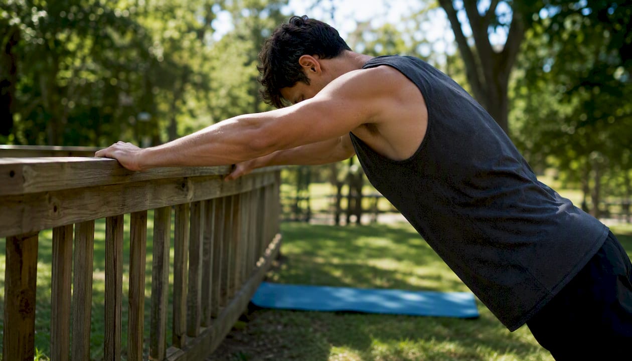
Early physical therapy to maintain range of motion and targeted strengthening is the initial mainstay of treatment and can prevent secondary complications such as adhesive capsulitis, which is a real risk when the shoulder is guarded and underused for months.
Therapy phases:
- Phase 1 — Activation and protection (weeks 1–8): Gentle range-of-motion work in pain-free planes. Supine shoulder flexion and gentle pendulum exercises preserve mobility without loading the denervated muscle. Scapular retraction and depression exercises activate the lower trapezius and rhomboids, which are often intact. Avoid aggressive overhead loading.
- Phase 2 — Endurance and strengthening (months 2–6+): As nerve recovery begins, introduce serratus anterior activation. The push-up plus generates high serratus anterior EMG activity and is the most commonly prescribed exercise for restoring scapular protraction and control. Closed-chain stabilization exercises (wall slides, quadruped reaches) build endurance without high joint loads.
- Phase 3 — Functional retraining (months 4–12+): Sport-specific or work-specific movements, progressive resistance, and overhead patterns introduced gradually as strength and control return.
Timeline expectations: Neuropraxic injuries commonly improve within several months. Clinicians typically observe recovery for a prolonged period before recommending surgery for nerve palsy. That’s a long time to wait, and patients need to understand it upfront.
Activity modification: Avoid repetitive overhead reaching, heavy pushing, and any activity that visibly worsens the winging. Return to sport or overhead work should be gradual and guided by objective strength testing, not just symptom resolution.
Bracing and taping: Scapular braces and kinesiology taping can reduce the visible deformity and offload the periscapular muscles during daily activities. Tolerability is the main limitation. Many patients find rigid braces uncomfortable for extended wear, and tape adherence varies with activity level and skin sensitivity. These are supportive tools, not treatments.
Pro Tip: Aggressive high-intensity strengthening too early after an acute nerve injury can aggravate neural irritation. The successful strategy balances a watchful wait for reinnervation with gentle, guided stabilization. If a new exercise causes sharp, shooting, or worsening neurologic pain, stop it and report back to your clinician.
Escalation criteria: If there’s no measurable improvement after 3–4 months of consistent therapy, or if weakness is progressing rather than plateauing, electrodiagnostic testing and specialist referral are warranted.
When surgery is the right answer for scapular winging
Surgery is not the first answer, but it’s the right answer in specific situations. The key is knowing which situation you’re in.
Indications for surgical consideration:
- Failed conservative care after an appropriate observation window (typically 12–24 months for nerve palsy)
- Complete nerve transection (confirmed on EMG or at exploration after penetrating or iatrogenic injury)
- Acute muscle avulsion where early repair is indicated
- Persistent functional deficit or pain that significantly limits daily activities despite adequate rehab
- Rapidly progressive weakness suggesting an evolving lesion
Common surgical options in plain language:
- Nerve decompression or repair: When the nerve is compressed or partially torn, releasing the compression or directly repairing the nerve can restore function. Best results come from early intervention after iatrogenic or penetrating injuries.
- Nerve transfer: A functioning donor nerve is rerouted to reinnervate the paralyzed muscle. Used when the original nerve cannot be repaired directly.
- Dynamic muscle transfer (pectoralis major transfer): For chronic serratus anterior palsy, the pectoralis major is rerouted to substitute for the lost serratus function. Outcomes in small case series are generally favorable, though patient selection and graft augmentation with fascia lata or hamstring tendon influence success rates.
- Eden-Lange procedure: For trapezius palsy, adjacent muscles (levator scapulae, rhomboids) are transferred to restore the trapezius’s stabilizing function. This is the standard dynamic transfer for lateral winging from spinal accessory nerve injury.
- Scapulothoracic fusion (scapulopexy): A salvage procedure that fuses the scapula to the rib cage, eliminating winging permanently but also eliminating scapulothoracic motion. Reserved for patients who have failed other options and prioritize stability over mobility.
Recovery after surgery varies by procedure. Nerve repairs and transfers require months for reinnervation to occur, often 6–18 months before meaningful strength returns. Muscle transfers typically involve 6–12 weeks of immobilization followed by structured rehabilitation. Scapulothoracic fusion has the longest recovery and most significant functional trade-off.
Surgical timing matters. For penetrating or iatrogenic nerve injuries, early exploration (within weeks) produces better outcomes than delayed repair. For long-standing palsy from neuropraxia that failed to recover, delayed muscle transfer is appropriate and can still produce good results.
What to expect: recovery timelines and prognosis
Most patients with neuropraxic scapular winging do recover, but the timeline is longer than most expect. Here’s a realistic picture:
| Recovery Scenario | Typical Timeline | Notes |
|---|---|---|
| Neuropraxia (nerve stretch/stun) | 6–9 months commonly | High recovery potential with conservative care |
| Conservative observation window | Up to 24 months | Standard before recommending surgery for nerve palsy |
| Surgical nerve repair/transfer | 6–18 months post-op | Depends on nerve regeneration rate |
| Dynamic muscle transfer | 6–12 months post-op | Includes immobilization and rehab phases |
| Scapulothoracic fusion | 12+ months | Significant functional trade-off for stability |
Factors that improve prognosis:
- Isolated serratus anterior palsy from a stretch or neuropraxic mechanism
- Younger age at injury
- Early initiation of range-of-motion preservation and guided rehab
- No secondary complications (adhesive capsulitis, rotator cuff involvement)
Factors that worsen prognosis:
- Complete nerve transection
- High-energy trauma with multiple structure involvement
- Delayed treatment beyond the optimal surgical window for acute injuries
- Age over 50 in some surgical series, associated with slower nerve regeneration
- Neuromuscular disease as the underlying cause (recovery is not expected; management focuses on function preservation)
Most patients with isolated neuropraxic winging can expect meaningful functional recovery, though some residual weakness or asymmetry may persist. The functional goal is restoring enough strength and control to return to daily activities and, for athletes, sport-specific demands. Full cosmetic resolution is common with complete nerve recovery but not guaranteed in all cases.
Prevention and red flags that need prompt attention
Not every case of scapular winging is preventable, but several risk factors are modifiable.
Prevention checklist:
- Use proper lifting technique: keep loads close to the body, avoid sudden jerking motions, and don’t let the shoulder blade “collapse” forward under load.
- Avoid extreme neck traction positions during contact sports, wrestling, or heavy stretching.
- Progress overhead sport training gradually, especially after a break or at the start of a new season.
- After neck or chest surgery, ask your surgeon specifically about protecting the spinal accessory and long thoracic nerves during recovery.
- Use properly fitted backpacks with padded, wide shoulder straps. Heavy single-strap bags worn across one shoulder are a documented risk for long thoracic nerve compression.
- Maintain scapular stabilizer strength as part of any shoulder conditioning program, particularly for overhead athletes. Posture correction and reducing neck/shoulder strain are part of a long-term prevention strategy.
Red flags requiring urgent evaluation (go to the ER or call your surgeon immediately):
- New difficulty breathing or swallowing alongside shoulder weakness
- Rapidly progressive arm or hand weakness over hours to days
- Signs of neck or vascular injury after trauma (bruising, pulsating mass, severe neck pain)
- Fever with acute neurologic change (possible infectious or inflammatory cause)
- Severe trauma to the chest, shoulder, or neck with immediate loss of function
First-aid steps for acute injuries: immobilize the arm in a sling if pain is severe, avoid all overhead use, apply ice for the first 48–72 hours, and document the exact mechanism of injury before your memory fades. That history is genuinely useful to the clinician.
Day-to-day rehab: safe home exercises and what to avoid
Home exercises are a complement to supervised physical therapy, not a replacement. That said, there are safe, useful things you can do between appointments.
Starter home program:
- Supine shoulder flexion (gravity-assisted range of motion): Lie on your back and use your unaffected arm to gently guide the affected arm overhead. This preserves range of motion without loading the weakened muscle. Stop at the point of discomfort.
- Scapular retraction squeezes: Sitting upright, gently squeeze the shoulder blades together and hold for 5 seconds. This activates the rhomboids and middle trapezius, which are often intact and can provide some compensatory support.
- Wall push-up plus (progressed carefully): Stand facing a wall, place both hands at shoulder height, and perform a slow push-up. At the top of the push, add a small extra push to protract the scapula (“plus” phase). This exercise generates high serratus anterior EMG activity and is the cornerstone of serratus reactivation. Start with 2–3 sets of 8–10 reps, and only progress if there’s no increase in neurologic symptoms.
- Gentle cervical range of motion: Slow, controlled neck movements (flexion, extension, rotation) help maintain mobility and reduce periscapular tension without stressing the injured nerve.
What to avoid at home:
- Aggressive overhead stretching or weighted overhead pressing before the nerve has recovered
- Resistance band exercises that load the serratus anterior at end-range before baseline activation is established
- Forcing through sharp or shooting pain, which may indicate nerve irritation rather than productive muscle fatigue
Pro Tip: The difference between productive discomfort and nerve irritation matters. Muscle fatigue feels like a dull burn that fades within minutes of stopping. Nerve irritation feels sharp, electric, or radiating, and it lingers. If your home exercises produce the second type, back off the intensity and discuss it with your therapist at the next visit.
Supervised physical therapy is essential for progressing beyond the starter phase. A therapist can observe your scapular mechanics in real time, adjust exercise selection based on what they see, and catch compensatory patterns before they become habits. Safe progressions for shoulder mobility are well-established in the literature, but applying them correctly to a nerve-injured shoulder requires clinical judgment.
How Thetxroom approaches scapular dysfunction
At Thetxroom in Plano, Texas. The approach to scapular winging starts with a thorough assessment: movement screening, manual palpation of the periscapular soft tissues, and a functional evaluation to identify which stabilizers are underperforming and whether any fascial restrictions are limiting normal scapulothoracic motion.
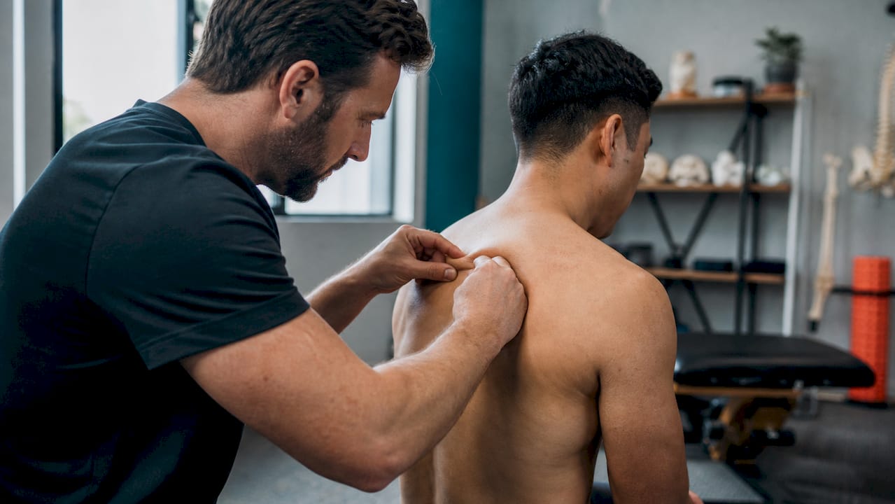
The primary treatment methodology is the Fascial Distortion Model (FDM), a hands-on approach that addresses soft-tissue distortions contributing to pain, restricted motion, and altered mechanics. For scapular dysfunction, this means working directly on the periscapular fascia, the serratus anterior, the trapezius, and the surrounding connective tissue to reduce tension, restore glide, and improve the quality of movement before progressive loading begins.
Early visits focus on range-of-motion restoration and soft-tissue desensitization. Mid-course care shifts toward targeted stabilization exercises, scapular motor control drills, and incremental home programs that patients can manage between sessions. Brace and taping guidance is provided where appropriate.
When red flags or surgical indications are present, Thetxroom coordinates transparently with orthopedic surgeons, neurologists, and other specialists. The clinic’s role is to optimize the patient’s condition before and after specialist intervention, not to delay necessary referrals.
For patients with shoulder pain who want hands-on care alongside their medical management, Thetxroom offers a practical, progressive path back to function.
A realistic perspective on recovery from scapular winging
The most common mistake I see is patients expecting a two-week fix for what is fundamentally a nerve-recovery problem. Nerves regenerate slowly, roughly 1 millimeter per day under ideal conditions, and the long thoracic nerve has a lot of ground to cover. That biological reality doesn’t change with more aggressive therapy or more frequent visits.
What does change outcomes is consistency: protecting the joint during the waiting period, maintaining range of motion so the shoulder doesn’t freeze up, and building stabilizer endurance so the muscle is ready to work when reinnervation arrives. The patients who do best are the ones who understand the timeline upfront and commit to the process without either panicking at slow progress or abandoning therapy when symptoms plateau.
The other thing worth saying plainly: most neuropraxic cases do recover. The conservative approach isn’t passive. It’s the evidence-based choice for the most common presentation, and it works for the majority of patients who follow through with it.
Thetxroom offers hands-on care for scapular winging in Plano, Texas
If you’re dealing with a winged shoulder blade and want hands-on treatment rather than a wait-and-see approach, Thetxroom in Plano, Texas, is built for exactly this kind of case. The clinic uses the Fascial Distortion Model to address the soft-tissue restrictions and biomechanical imbalances that accompany scapular dysfunction, combined with targeted stabilization work and progressive rehab plans tailored to where you are in recovery.

Thetxroom works with athletes, desk workers, post-surgical patients, and anyone dealing with periscapular pain or weakness who wants a clear plan and measurable progress. When surgical or neurological referral is warranted, the clinic coordinates that directly rather than leaving patients to navigate it alone.
To get started, complete your new patient paperwork online and book your initial evaluation. You’ll leave the first visit with a working diagnosis, a clear explanation of what’s happening, and a treatment plan you can act on immediately.
Sources
The following sources were used throughout this article and are worth bookmarking for further reference:
- Winging of the Scapula – StatPearls – NCBI Bookshelf
- Winged Scapula: Causes, Symptoms, Tests & Treatment
- Scapular winging: anatomical review, diagnosis, and treatments
- Scapular Winging – Shoulder & Elbow
- Scapular winging: anatomical review, diagnosis, and treatments
This article is general information, not a substitute for advice from a qualified doctor. Consult a qualified healthcare professional about your own circumstances before acting on anything here.