

Pinched Nerve Neck Relief: Safe Exercises & Home Plan
Targeted self-care and specific neck exercises usually reduce pinched nerve neck pain within days to weeks. Start with ice for the first 48–72 hours, then switch to heat, modify activities to avoid positions that provoke symptoms, and add gentle isometric holds and nerve glides once they ease your pain rather than worsen it. About 85% of cervical radiculopathy cases resolve or greatly improve within 8–12 weeks with conservative care alone.
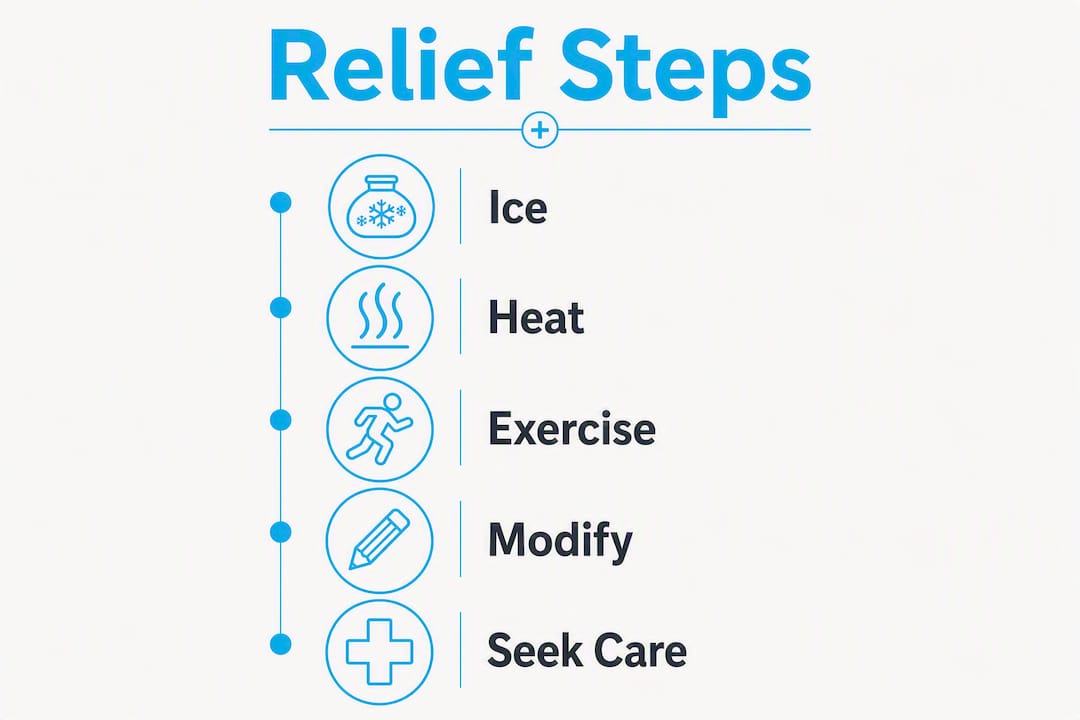
The short version of what to do right now:
- Apply ice (wrapped in a cloth) to the neck for 15–20 minutes, up to several times a day for the first 48–72 hours.
- After 72 hours, switch to a heating pad or warm shower to relax tight muscles.
- Take an OTC NSAID like ibuprofen or naproxen as directed on the label if you tolerate them.
- Modify activity: avoid heavy lifting, overhead reaching, or any posture that loads the neck.
- Begin gentle isometric holds and nerve glides only if they reduce, not increase, your symptoms.
One rule that overrides everything else: stop any stretch or movement the moment it increases radiating pain into your arm or hand. That spike means the inflamed nerve root is being irritated further. Gentle exercises can speed recovery, but only when they do not provoke nerve pain.
Seek care sooner if you notice progressive arm weakness, new numbness spreading into your hand, or any change in bowel or bladder function. Those signs move this from a “wait and manage” situation to a “call today” one. Most cases are managed without surgery, but the 6–12 week conservative window is the standard clinical benchmark before surgical options are discussed.
Table of Contents
- What causes a pinched nerve in your neck?
- How a pinched nerve in the neck typically feels
- How clinicians diagnose a pinched nerve in the neck
- Your step-by-step home plan for immediate relief
- The best exercises for a pinched nerve in the neck
- When to consider professional treatment for your neck
- Medical treatments and surgical options
- When to see a doctor: red flags and timing
- What to expect during recovery
- How to prevent a pinched nerve from coming back
- The hands-on approach to treating cervical nerve compression
- Key Takeaways
- The part most people get wrong about neck nerve recovery
- Thetxroom in Plano can help you recover faster
- Useful sources and further reading
What causes a pinched nerve in your neck?
The clinical term is cervical radiculopathy: compression or inflammation of a nerve root as it exits the cervical spine, producing pain, tingling, numbness, or weakness that can radiate from the neck into the shoulder, arm, or hand. The nerve root itself is not always physically “pinched” in the dramatic sense; sometimes it is simply inflamed from nearby tissue pressing against it.
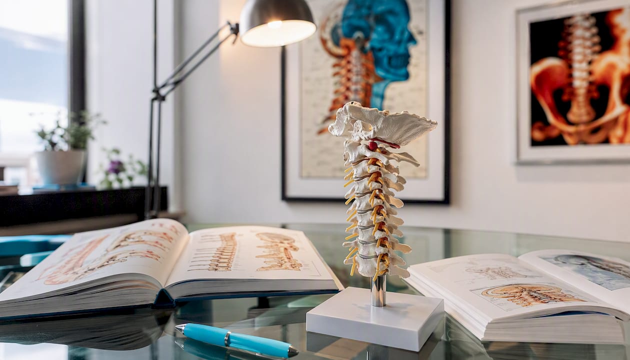
Age-related degenerative changes account for an estimated 70–90% of cases, which is why cervical radiculopathy is more common after 40 and why long-term biomechanical maintenance matters as much as acute treatment. The spine gradually loses disc height and develops bone spurs (osteophytes) that narrow the foraminal openings where nerve roots exit.
Common causes:
- Degenerative disc disease: discs lose hydration and height over time, reducing the space available for nerve roots.
- Herniated or bulging disc: the inner disc material pushes outward and presses on an adjacent nerve root, often from a sudden load or twisting movement.
- Bone spurs (osteophytes): bony growths that form at disc margins or facet joints can directly narrow the foramen.
- Trauma or whiplash: a sudden force, such as a car accident or sports collision, can shift disc material or strain supporting structures enough to compress a nerve root. Whiplash-related cervical injuries are a common presentation in clinical practice.
- Poor posture and repetitive strain: sustained forward-head posture (common with desk work and phone use) loads the lower cervical segments unevenly, accelerating degenerative change and increasing nerve root stress over time.
How a pinched nerve in the neck typically feels
Cervical radiculopathy has a recognizable symptom pattern that distinguishes it from ordinary neck stiffness. The hallmark is that symptoms travel. General neck strain stays local; radiculopathy sends signals down the nerve’s distribution path.
Typical symptoms:
- Sharp, shooting, or burning neck pain that may radiate into the shoulder, upper arm, forearm, or hand.
- Tingling or “pins and needles” along the arm or into specific fingers (the exact fingers depend on which nerve root is involved).
- Numbness in the arm or hand, sometimes described as the limb “falling asleep.”
- Muscle weakness in the shoulder, arm, or grip, which can be subtle at first.
- Reduced reflexes at the biceps, triceps, or brachioradialis, detectable on clinical exam.
- Neck stiffness and reduced range of motion, particularly with rotation or side-bending toward the affected side.
Symptoms often worsen when you extend or rotate the neck toward the affected side, and many people find relief by raising the arm overhead (which reduces tension on the nerve root).
Red flags that need immediate evaluation:
- Progressive motor weakness (dropping objects, difficulty raising your arm).
- Loss of bowel or bladder control.
- Fever combined with neck pain and stiffness.
- Severe, unremitting pain that does not respond to any position change.
- Sudden inability to use an arm or walk normally.
These signs suggest something beyond a typical pinched nerve and require same-day or emergency assessment.
How clinicians diagnose a pinched nerve in the neck
Diagnosis follows a logical sequence, and imaging is not always the first step. A skilled clinician can often identify the likely nerve root involved from the physical exam alone, which guides whether and what imaging is needed.
-
Focused neurological and physical exam. The clinician tests neck range of motion, upper-extremity strength (grip, wrist extension, shoulder abduction), sensation in dermatomal patterns, and deep tendon reflexes. The Spurling test, where the neck is extended and rotated toward the symptomatic side with gentle downward pressure, reproduces radicular symptoms when positive and is one of the most specific clinical tests for cervical radiculopathy.
-
X-ray. Ordered to assess bony alignment, disc space height, and the presence of osteophytes. X-ray does not show soft tissue or nerve roots directly, but it reveals degenerative changes and rules out fractures or instability.
-
MRI. The preferred imaging for visualizing disc herniation, nerve root compression, spinal cord involvement, and soft-tissue detail. MRI is the standard when symptoms are severe, progressive, or not responding to initial conservative care, or when surgery is being considered.
-
CT scan. Used when MRI is contraindicated (pacemaker, certain implants) or when bony detail is the primary concern. CT myelography, where contrast dye is injected into the spinal canal before scanning, provides excellent nerve root visualization when MRI is unavailable.
-
Electromyography (EMG) and nerve conduction studies (NCS). These tests measure the electrical activity of muscles and the speed of nerve signals. They are not ordered routinely; clinicians use them when the diagnosis is uncertain, when symptoms have persisted for several weeks, or when the clinical exam does not clearly localize the affected nerve root. EMG can confirm nerve root dysfunction and distinguish radiculopathy from peripheral nerve entrapment (like carpal tunnel syndrome).
The key point: most people with a first episode of cervical radiculopathy start conservative care based on the clinical exam, without waiting for imaging results. Imaging is reserved for cases that are severe, atypical, or not improving.
Your step-by-step home plan for immediate relief
The goal in the first few days is to reduce inflammation, protect the nerve root from further irritation, and keep moving without provoking symptoms. Total bed rest is outdated advice; staying as active as possible while avoiding provoking movements speeds recovery and prevents deconditioning.
Days 1–3 (acute phase):
- Ice first. Apply ice for the first 48–72 hours to reduce inflammation and acute pain. Wrap a cold pack in a thin cloth and apply to the neck for 15–20 minutes at a time, several times daily. Never apply ice directly to skin.
- Modify activity. Avoid heavy lifting, overhead work, and any posture that loads the neck forward or backward. Driving long distances, looking down at a phone, and sleeping on your stomach are common aggravators.
- OTC medication. Ibuprofen (Advil, Motrin) or naproxen (Aleve) reduce both pain and inflammation. Acetaminophen (Tylenol) addresses pain but not inflammation. Take as directed and consult a pharmacist if you have kidney, stomach, or cardiovascular concerns.
- Sleep position. Sleep on your back or side with a supportive pillow that keeps the neck in neutral alignment. A cervical contour pillow or a rolled towel inside a pillowcase can help. Avoid sleeping on your stomach, which rotates and extends the neck.
Days 3 onward:
- Switch to heat. After the first 48–72 hours, heat relaxes muscles and improves flexibility. A warm shower, heating pad on low, or warm towel applied for 15–20 minutes before gentle movement works well.
- Soft collar use. A soft cervical collar can provide short-term relief by limiting painful movements, but limit use to a few hours per day and no more than 1–2 weeks. Prolonged collar use weakens neck muscles and slows recovery.
- Prescription options. If OTC medications are not controlling pain, a clinician may prescribe a short course of oral corticosteroids to reduce nerve root inflammation, or a muscle relaxant for severe spasm. NSAIDs are the common first-line option; oral corticosteroids are reserved for severe, persistent nerve-related inflammation.
When home care is not enough: contact a clinician if pain is not improving after 1–2 weeks of consistent self-care, if radiating symptoms are spreading or worsening, or if you notice any weakness in the arm or hand.
The best exercises for a pinched nerve in the neck

Warm up before any neck exercise: a warm shower or 10 minutes with a heating pad loosens the tissue and makes movement safer. Work through these daily, and stop immediately if any movement increases radiating arm pain.
The non-negotiable rule: if a movement sends pain, tingling, or numbness further down your arm, stop. That is nerve irritation, not a “good stretch.”
-
Isometric neck holds (start here for acute pain)
Purpose: Activate the deep cervical stabilizers without moving the neck, reducing muscle spasm while protecting the nerve root.
How: Sit upright. Place your palm against your forehead. Press your head gently into your hand without letting either move. Hold 5–10 seconds. Repeat on each side (palm against temple) and at the back of the head. 3 sets of 5 reps per direction. Isometric exercises are the safest starting point in acute pain because they support the neck without risking excessive movement. -
Chin tuck (cervical retraction)
Purpose: Corrects forward-head posture and reduces load on the lower cervical nerve roots.
How: Sit or stand tall. Gently draw your chin straight back (not down) as if making a “double chin.” Hold 3–5 seconds, then release. 10 reps, 2–3 times daily. If this reproduces arm symptoms, reduce the range or skip it until symptoms settle. -
Ear-to-shoulder stretch (lateral flexion)
Purpose: Lengthens the scalene and upper trapezius muscles that often tighten around an irritated nerve root.
How: Sit upright. Slowly tilt your right ear toward your right shoulder until you feel a gentle stretch on the left side of the neck. Hold 20–30 seconds. Return to center. Repeat on the other side. 3 reps per side. Never force the stretch or use your hand to pull the head down. -
Neck rotation
Purpose: Restores rotational mobility and reduces stiffness without loading the nerve root.
How: Sit tall. Slowly turn your head to the right as far as comfortable, pause 2–3 seconds, return to center, then turn left. 5–10 reps per side. Move only within a pain-free range. -
Shoulder rolls and scapular retraction
Purpose: Releases upper trapezius tension and activates the mid-back muscles that support cervical posture.
How: Roll shoulders backward in slow, large circles, 10 reps. Then squeeze shoulder blades together and hold 5 seconds, 10 reps. These are safe even in acute phases and directly reduce the postural load on the neck. -
Cervical nerve glide (median nerve bias)
Purpose: Mobilizes the nerve root and its surrounding tissue to reduce adhesion and improve neural mobility.
How: Sit upright. Tilt your head away from the symptomatic arm (right ear toward right shoulder if the left arm is affected). Simultaneously extend the symptomatic arm out to the side with the wrist bent back (palm facing away). Hold 3–5 seconds, then return to neutral. 5–10 slow reps. This should produce a mild stretch sensation, never a sharp pain or increase in radiating symptoms. For a full guide on safe stretching protocols, see Thetxroom’s stretching guidance. -
Upper trapezius and levator scapulae stretch
Purpose: Targets the muscles most commonly in spasm with cervical radiculopathy.
How: Sit on your right hand (to anchor the shoulder). With your left hand, gently guide your head toward the left shoulder and slightly forward. Hold 30 seconds. Switch sides. 2–3 reps per side.
Pro Tip: Do these exercises after heat, not cold. Warm tissue is more pliable and responds better to gentle movement. If you do them first thing in the morning, spend 5 minutes in a warm shower before starting.
When to consider professional treatment for your neck
Home care handles most mild to moderate cases, but there are clear signals that professional hands-on care will get you further, faster. Most mild neck pain improves within 2–3 weeks with self-care; cervical radiculopathy often needs up to 8–12 weeks of structured conservative management.
Signs it is time to see a professional:
- Symptoms are not improving after 1–2 weeks of consistent home care.
- Radiating pain, numbness, or tingling is spreading or intensifying.
- You have functional limitations at work, driving, or during daily tasks.
- You cannot sleep due to pain despite position modifications.
- You have had a prior episode and recognize the pattern escalating.
What professional options look like:
- Physical therapy: A physical therapist designs a progressive exercise program, may use modalities like ultrasound or electrical stimulation, and addresses postural and ergonomic contributors. The focus is on active rehabilitation and building the strength to prevent recurrence.
- Chiropractic and manual therapy: Chiropractors and manual therapists address joint mobility restrictions and soft-tissue tension around the affected segments. Techniques range from gentle mobilization to specific soft-tissue work. Chiropractic care for neck conditions focuses on restoring biomechanical function, not just symptom suppression.
- Cervical traction: Mechanical or manual traction gently distracts the cervical segments, increasing foraminal space and reducing nerve root compression. It is most effective for disc-related radiculopathy and is typically applied by a trained clinician or with a home traction device prescribed by one.
Choosing a provider: ask whether they have experience treating cervical radiculopathy specifically, what outcome measures they use to track progress, and what their safety precautions are for forceful techniques. A good clinician sets clear goals and adjusts the plan based on your response, not a fixed protocol.
Medical treatments and surgical options
| Treatment | Typical indication | Expected effect | Key notes |
|---|---|---|---|
| OTC NSAIDs (ibuprofen, naproxen) | Mild to moderate pain and inflammation | Reduces pain and swelling within days | Take with food; avoid with kidney/GI issues |
| Acetaminophen | Pain without significant inflammation | Analgesic only; no anti-inflammatory effect | Safer for those who cannot tolerate NSAIDs |
| Muscle relaxants (cyclobenzaprine, etc.) | Significant muscle spasm | Reduces spasm; sedating | Short-term use; prescription required |
| Nerve pain medications (gabapentin, pregabalin) | Persistent neuropathic/radiating pain | Reduces nerve-mediated pain signals | Prescription; may cause drowsiness |
| Oral corticosteroids (prednisone) | Severe nerve root inflammation | Rapid reduction of inflammation | Short course only; not for long-term use |
| Epidural steroid injection (ESI) | Persistent radiculopathy not responding to oral meds | Targeted anti-inflammatory effect at nerve root | Performed by pain specialist; effects vary |
| Surgery (ACDF, disc replacement) | Pain >6–12 weeks despite conservative care, or worsening neurologic deficit | Decompresses nerve root; relieves radicular symptoms | Reserved for specific indications; realistic recovery is weeks to months |
Surgery is not the default outcome. Most cervical radiculopathy cases are managed without surgery, and the decision to operate is based on a combination of symptom duration, severity of neurological deficit, and failure of structured conservative care. When surgery is indicated, the most common procedure is anterior cervical discectomy and fusion (ACDF), which removes the offending disc material and stabilizes the segment. Disc replacement is an alternative that preserves motion at the treated level.
Epidural steroid injections sit between oral medications and surgery in the treatment ladder. They deliver corticosteroid directly to the epidural space near the affected nerve root, providing targeted relief that can allow a patient to participate more fully in physical rehabilitation. Effects are temporary in many cases, but they can be a meaningful bridge.
When to see a doctor: red flags and timing
Seek immediate (same-day or emergency) care for:
- Progressive motor weakness, such as inability to grip, raise your arm, or lift your wrist.
- Loss of bowel or bladder control.
- Sudden, severe neck pain after trauma (fall, accident, impact).
- Fever with neck pain and stiffness (possible meningitis or infection).
- Sudden inability to walk normally or use an arm.
Seek prompt care within a few days for:
- Severe radiating pain that is not responding to OTC medication and position changes.
- Numbness or tingling that is spreading or worsening over 24–48 hours.
- Symptoms that began after a car accident or sports injury.
Schedule a routine appointment if:
- Symptoms are stable but not improving after 1–2 weeks of consistent home care.
- This is a recurrent episode and you want a formal assessment and structured plan.
When you go, bring a brief symptom log: when it started, what makes it worse or better, which arm or fingers are affected, and any prior imaging you have. That information cuts the diagnostic time significantly and helps the clinician order the right tests on the first visit.
What to expect during recovery
Recovery from cervical radiculopathy is not linear, but the overall trajectory for most people is positive. The first two weeks typically bring the most noticeable fluctuation: some days feel better, others worse, often tied to sleep quality, activity level, and posture.
| Timeframe | Typical experience |
|---|---|
| Days 1–7 | Acute pain and muscle spasm; focus on ice/heat, activity modification, and OTC medication |
| Weeks 2–4 | Gradual reduction in resting pain; begin gentle exercises if tolerated; radiating symptoms may persist |
| Weeks 4–8 | Meaningful improvement in most cases; progressive exercise and manual therapy if needed |
| Weeks 8–12 | Most cases resolve or substantially improve; reassess if symptoms remain significant |
| Beyond 12 weeks | Persistent or worsening symptoms warrant imaging, specialist referral, or reconsideration of treatment approach |
About 85% of cervical radiculopathy cases resolve or greatly improve within 8–12 weeks. That figure is genuinely reassuring, but it does not mean passive waiting. The cases that do best are the ones where people stay active, follow a structured exercise plan, and address the biomechanical contributors rather than just managing symptoms.
Factors that affect how quickly you recover:
- Age and degree of degenerative change: more advanced degeneration typically means a longer recovery.
- Smoking: reduces disc nutrition and impairs tissue healing.
- Activity level: people who stay active within their tolerance recover faster than those who rest completely.
- Promptness of appropriate care: starting structured rehabilitation early, rather than waiting weeks in pain, consistently produces better outcomes.
- Psychological factors: high pain catastrophizing and poor sleep are associated with slower recovery, independent of the physical severity.
How to prevent a pinched nerve from coming back
Most recurrences are preventable with consistent attention to posture, ergonomics, and maintenance exercise. The same degenerative process that caused the first episode continues unless you actively counteract it.
Ergonomics checklist:
- Monitor at eye level so the neck stays neutral, not flexed forward.
- Chair back supports the lumbar spine; feet flat on the floor or on a footrest.
- Elbows at roughly 90 degrees when typing; keyboard and mouse close to the body.
- Take a standing or walking break every 30–45 minutes during desk work.
- Hold the phone at eye level rather than looking down; use a headset for long calls.
- When lifting, hinge at the hips and keep the load close to the body; avoid twisting while loaded.
Maintenance exercises to do daily:
- Chin tucks: 10 reps, 2–3 times daily (the single most effective posture-correction exercise for the cervical spine).
- Scapular retractions: 10–15 reps to maintain mid-back activation and reduce upper trapezius overload.
- Thoracic extension over a foam roller: 1–2 minutes to counteract the thoracic flexion that drives forward-head posture.
- Graded cervical strengthening: as symptoms resolve, progress to resistance band exercises for the deep neck flexors and extensors.
For athletes and active individuals, a structured mobility restoration program that includes cervical and thoracic mobility work is one of the most effective ways to reduce recurrence risk.
Lifestyle factors that matter:
- Maintaining a healthy weight reduces axial load on the cervical spine.
- Quitting smoking improves disc nutrition and tissue repair capacity.
- Regular aerobic activity supports spinal health and reduces systemic inflammation.
- If symptoms recur, schedule a professional check-in early rather than waiting for them to escalate.
The hands-on approach to treating cervical nerve compression
Generic stretching addresses muscle tightness. What it often misses is the fascial and soft-tissue restriction that builds up around a chronically irritated nerve root, limiting its ability to glide freely and contributing to repeated flare-ups. This is where manual therapy adds something that home exercises alone cannot replicate.
At Thetxroom, the primary hands-on framework is the Fascial Distortion Model (FDM), a diagnostic and treatment approach that identifies specific types of fascial distortion contributing to pain and restricted movement. Rather than applying a generic protocol, FDM-trained practitioners assess how the patient describes and gestures toward their pain, using that information to identify the tissue-level problem and apply targeted manual correction.
What this approach addresses beyond standard stretching:
- Fascial bands and trigger points that restrict nerve root mobility.
- Joint mobility restrictions in the cervical and upper thoracic segments that increase load on specific nerve roots.
- Soft-tissue tension patterns that perpetuate postural imbalances even after pain subsides.
- Biomechanical contributors in the shoulder and thoracic spine that refer strain upward into the neck.
The clinic integrates manual therapy with progressive exercise and a tailored home plan, so patients leave each session with something to do between visits, not just passive treatment. For range-of-motion restoration specifically, combining hands-on work with active exercise consistently produces more durable improvement than either alone.
How to choose a manual therapist for cervical radiculopathy:
- Confirm they have specific experience treating cervical radiculopathy, not just general neck pain.
- Ask what outcome measures they use to track progress (range of motion, pain scores, functional tasks).
- Expect a clear explanation of what they are treating and why, not just “adjustments.”
- A good therapist adjusts the plan based on your response between sessions.
Pro Tip: Between visits, keep a brief log: rate your pain (0–10), note which activities aggravated or relieved it, and track whether radiating symptoms are changing. That data helps your clinician adjust the treatment plan far more precisely than a verbal summary.
Key Takeaways
Most pinched nerve neck pain resolves within 8–12 weeks with consistent conservative care: ice then heat, activity modification, safe exercises, and professional hands-on treatment when home care is not enough.
| Point | Details |
|---|---|
| Start with ice, then heat | Ice for the first 48–72 hours reduces inflammation; switch to heat after that to relax muscles. |
| Stop if nerve pain increases | Any exercise or movement that worsens radiating arm pain should be stopped immediately. |
| 85% resolve without surgery | About 85% of cervical radiculopathy cases greatly improve within 8–12 weeks of conservative care. |
| See a doctor for red flags | Progressive weakness, loss of bowel/bladder control, or severe unremitting pain need prompt evaluation. |
| Thetxroom offers hands-on care | FDM-based manual therapy at Thetxroom in Plano addresses soft-tissue and biomechanical contributors that home exercises miss. |
The part most people get wrong about neck nerve recovery
There is a persistent belief that if something hurts, you should rest it completely. For a broken bone, that is correct. For a pinched nerve in the neck, it is usually the wrong call. The research is clear: staying active within your tolerance produces better outcomes than bed rest, and the patients who do best are the ones who start structured movement early, not the ones who wait for the pain to disappear before doing anything.
The second mistake is treating this as purely a structural problem. Yes, a herniated disc or bone spur is the mechanical trigger. But the reason some people recover in three weeks and others are still struggling at three months often has less to do with the disc and more to do with the surrounding tissue: the fascial restrictions, the postural habits, the muscle imbalances that were there before the nerve got irritated and will still be there after the acute pain fades. Address only the acute symptoms and you are setting up the next episode.
The third thing people underestimate is the upper thoracic spine. The cervical spine does not work in isolation. Stiffness in the mid-back forces the neck to compensate, loading the same segments repeatedly. Thoracic mobility work is not glamorous, but it is one of the most reliable ways to reduce recurrence. Most home exercise programs for neck pain skip it entirely.
If you are in the early days of a pinched nerve flare, the practical takeaway is this: ice it, move gently, do not catastrophize, and get a proper assessment if it is not improving in two weeks. The odds are strongly in your favor.
Thetxroom in Plano can help you recover faster
Neck nerve compression that is not responding to home care usually has a mechanical contributor that self-treatment cannot fully address. At Thetxroom in Plano, Texas, the first visit includes a focused movement and neurological assessment, hands-on soft-tissue work using the Fascial Distortion Model, and a tailored home plan you can start the same day.

The FDM approach targets the specific fascial and joint restrictions driving your symptoms, not a generic protocol. Most patients notice a measurable change in range of motion and pain intensity within the first few sessions. For cases rooted in disc-related compression, the clinic combines manual therapy with progressive rehabilitation to address both the acute nerve irritation and the biomechanical pattern that caused it.
Ready to stop managing symptoms and start resolving them? Complete your new patient paperwork and book your first visit at Thetxroom today.
Useful sources and further reading
The following sources were used to support the clinical claims, timelines, and treatment guidance in this article:
- Harvard Health: Treating a Pinched Nerve — Source for the 70–90% degenerative causation figure and long-term maintenance rationale.
- Harvard Health: How to Soothe a Sore Neck — Supports the ice-then-heat protocol and general self-care sequence.
- Harvard Health: Got a Pinched Nerve? Strategies and Treatments — Backs the exercise safety rule (stop if nerve pain increases) and the role of gentle movement in recovery.
- MedlinePlus: Activity Guidance for Acute Neck Pain — Supports the recommendation to stay active rather than rest completely.
- NINDS: Pinched Nerve Information Page — National Institute of Neurological Disorders and Stroke overview of nerve compression conditions.
- NCBI: Cervical Radiculopathy Review — Peer-reviewed clinical review covering pathophysiology, diagnosis, and treatment evidence.
This article provides general health information and is not a substitute for professional medical advice. Confirm current treatment recommendations and your specific situation with a licensed clinician.